Department of Neuroimmunology, Center for Brain Research, Medical University of Vienna, Vienna, Austria.
Ann Neurol. 2013 Dec;74(6):848-61. doi: 10.1002/ana.23974. Epub 2013 Oct 7.
Iron may contribute to the pathogenesis and progression of multiple sclerosis (MS) due to its accumulation in the human brain with age. Our study focused on nonheme iron distribution and the expression of the iron-related proteins ferritin, hephaestin, and ceruloplasmin in relation to oxidative damage in the brain tissue of 33 MS and 30 control cases.
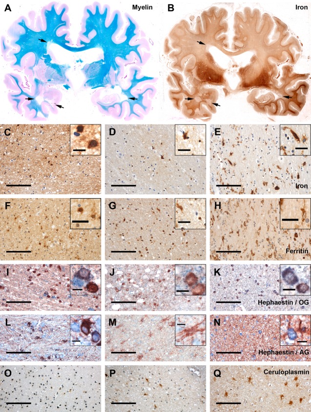
We performed (1) whole-genome microarrays including 4 MS and 3 control cases to analyze the expression of iron-related genes, (2) nonheme iron histochemistry, (3) immunohistochemistry for proteins of iron metabolism, and (4) quantitative analysis by digital densitometry and cell counting in regions representing different stages of lesion maturation.
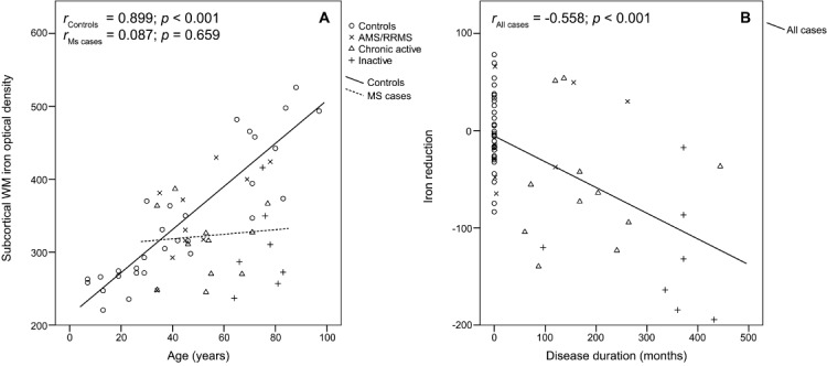
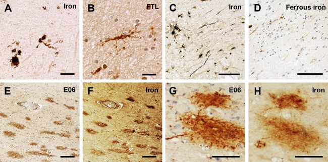
We found an age-related increase of iron in the white matter of controls as well as in patients with short disease duration. In chronic MS, however, there was a significant decrease of iron in the normal-appearing white matter (NAWM) corresponding with disease duration, when corrected for age. This decrease of iron in oligodendrocytes and myelin was associated with an upregulation of iron-exporting ferroxidases. In active MS lesions, iron was apparently released from dying oligodendrocytes, resulting in extracellular accumulation of iron and uptake into microglia and macrophages. Iron-containing microglia showed signs of cell degeneration. At lesion edges and within centers of lesions, iron accumulated in astrocytes and axons.
Iron decreases in the NAWM of MS patients with increasing disease duration. Cellular degeneration in MS lesions leads to waves of iron liberation, which may propagate neurodegeneration together with inflammatory oxidative burst.
由于铁在人体内的积累随年龄增长而增加,因此铁可能会导致多发性硬化症(MS)的发病和进展。我们的研究重点是研究非血红素铁分布以及铁相关蛋白铁蛋白、hephaestin 和铜蓝蛋白在 33 例 MS 和 30 例对照病例的脑组织中与氧化损伤的关系。
我们进行了(1)全基因组微阵列分析,包括 4 例 MS 和 3 例对照病例,以分析铁相关基因的表达;(2)非血红素铁组织化学;(3)铁代谢蛋白的免疫组织化学;(4)通过数字密度计量和细胞计数在代表病变成熟不同阶段的区域进行定量分析。
我们发现对照组和病程较短的患者的白质中存在与年龄相关的铁增加。然而,在慢性 MS 中,与年龄相关的正常外观白质(NAWM)中的铁含量随着疾病持续时间的延长而显著减少。当校正年龄时,铁在少突胶质细胞和髓鞘中的减少与铁输出铁氧化酶的上调有关。在活跃的 MS 病变中,铁显然从死亡的少突胶质细胞中释放出来,导致细胞外铁的积累并被小胶质细胞和巨噬细胞摄取。含铁血素的小胶质细胞显示出细胞退化的迹象。在病变边缘和病变中心内,铁在星形胶质细胞和轴突中积累。
随着疾病持续时间的增加,MS 患者的 NAWM 中铁含量减少。MS 病变中的细胞退化导致铁的释放波,这可能与炎症性氧化爆发一起传播神经退行性变。