Dal-Bianco Assunta, Grabner Günther, Kronnerwetter Claudia, Weber Michael, Höftberger Romana, Berger Thomas, Auff Eduard, Leutmezer Fritz, Trattnig Siegfried, Lassmann Hans, Bagnato Francesca, Hametner Simon
Department of Neurology, Medical University of Vienna, Vienna, Austria.
Department of Health Sciences and Social Work, Carinthia University of Applied Sciences, Klagenfurt, Austria.
Acta Neuropathol. 2017 Jan;133(1):25-42. doi: 10.1007/s00401-016-1636-z. Epub 2016 Oct 27.
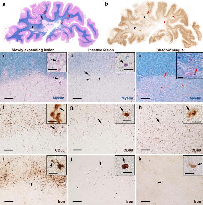
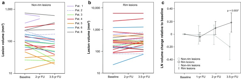
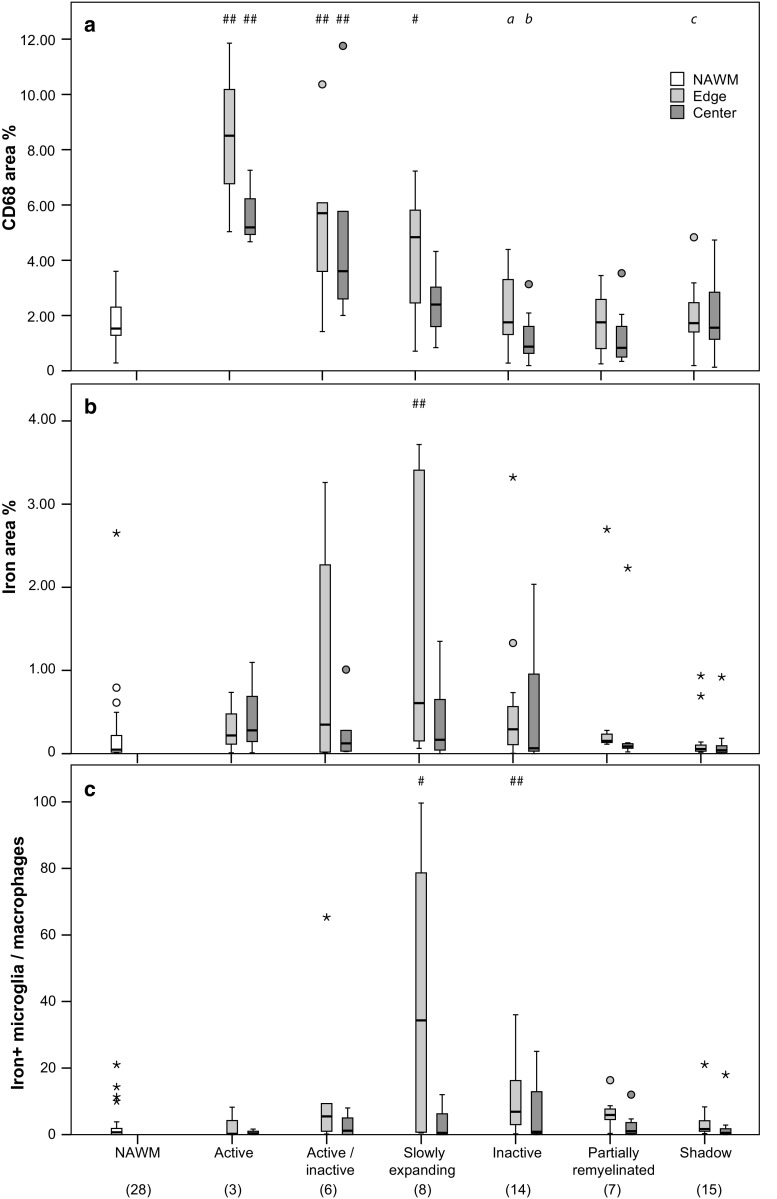
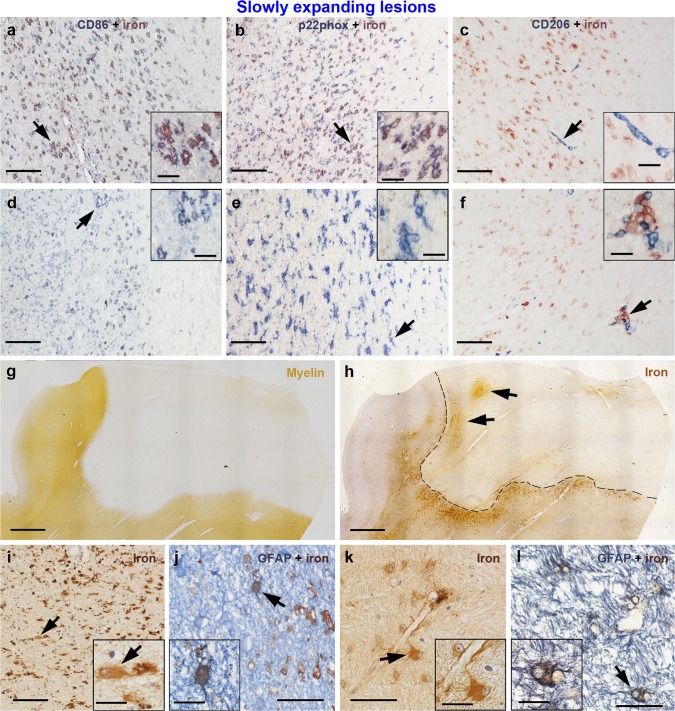
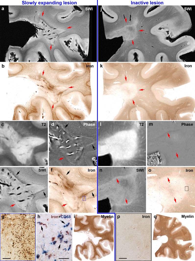
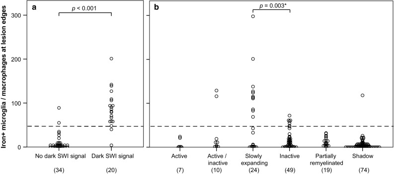
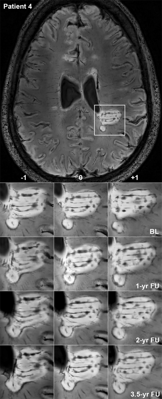
In multiple sclerosis (MS), iron accumulates inside activated microglia/macrophages at edges of some chronic demyelinated lesions, forming rims. In susceptibility-based magnetic resonance imaging at 7 T, iron-laden microglia/macrophages induce a rim of decreased signal at lesion edges and have been associated with slowly expanding lesions. We aimed to determine (1) what lesion types and stages are associated with iron accumulation at their edges, (2) what cells at the lesion edges accumulate iron and what is their activation status, (3) how reliably can iron accumulation at the lesion edge be detected by 7 T magnetic resonance imaging (MRI), and (4) if lesions with rims enlarge over time in vivo, when compared to lesions without rims. Double-hemispheric brain sections of 28 MS cases were stained for iron, myelin, and microglia/macrophages. Prior to histology, 4 of these 28 cases were imaged at 7 T using post-mortem susceptibility-weighted imaging. In vivo, seven MS patients underwent annual neurological examinations and 7 T MRI for 3.5 years, using a fluid attenuated inversion recovery/susceptibility-weighted imaging fusion sequence. Pathologically, we found iron rims around slowly expanding and some inactive lesions but hardly around remyelinated shadow plaques. Iron in rims was mainly present in microglia/macrophages with a pro-inflammatory activation status, but only very rarely in astrocytes. Histological validation of post-mortem susceptibility-weighted imaging revealed a quantitative threshold of iron-laden microglia when a rim was visible. Slowly expanding lesions significantly exceeded this threshold, when compared with inactive lesions (p = 0.003). We show for the first time that rim lesions significantly expanded in vivo after 3.5 years, compared to lesions without rims (p = 0.003). Thus, slow expansion of MS lesions with rims, which reflects chronic lesion activity, may, in the future, become an MRI marker for disease activity in MS.
在多发性硬化症(MS)中,铁在一些慢性脱髓鞘病变边缘的活化小胶质细胞/巨噬细胞内蓄积,形成环状物。在7T基于敏感性的磁共振成像中,富含铁的小胶质细胞/巨噬细胞在病变边缘诱导出信号降低的环状物,并与缓慢扩大的病变相关。我们旨在确定:(1)哪些病变类型和阶段与边缘铁蓄积相关;(2)病变边缘哪些细胞蓄积铁以及它们的活化状态如何;(3)7T磁共振成像(MRI)检测病变边缘铁蓄积的可靠性如何;(4)与无环状物的病变相比,有环状物的病变在体内随时间是否会扩大。对28例MS病例的双侧大脑切片进行铁、髓鞘和小胶质细胞/巨噬细胞染色。在组织学检查之前,这28例病例中的4例在死后使用敏感性加权成像在7T下成像。在体内,7例MS患者进行了为期3.5年的年度神经学检查和7T MRI检查,使用液体衰减反转恢复/敏感性加权成像融合序列。在病理上,我们发现在缓慢扩大的病变和一些静止性病变周围有铁环,但在再髓鞘化的阴影斑块周围几乎没有。环中的铁主要存在于具有促炎活化状态的小胶质细胞/巨噬细胞中,但在星形胶质细胞中很少见。死后敏感性加权成像的组织学验证显示,当可见环状物时,富含铁的小胶质细胞有一个定量阈值。与静止性病变相比,缓慢扩大的病变显著超过该阈值(p = 0.003)。我们首次表明,与无环状物的病变相比,有环状物的病变在3.5年后在体内显著扩大(p = 0.003)。因此,反映慢性病变活动的有环状物的MS病变的缓慢扩大,未来可能成为MS疾病活动的MRI标志物。