Department of Psychiatry, Medical University of South Carolina, Charleston, South Carolina, United States of America.
PLoS One. 2013 Jul 9;8(7):e67917. doi: 10.1371/journal.pone.0067917. Print 2013.
The prefrontal cortex (PFC) is an anatomically and functionally heterogeneous area which influences cognitive and limbic processing through connectivity to subcortical targets. As proposed by Alexander et al. (1986) the lateral and medial aspects of the PFC project to distinct areas of the striatum in parallel but functionally distinct circuits. The purpose of this preliminary study was to determine if we could differentially and consistently activate these lateral and medial cortical-subcortical circuits involved in executive and limbic processing though interleaved transcranial magnetic stimulation (TMS) in the MR environment.
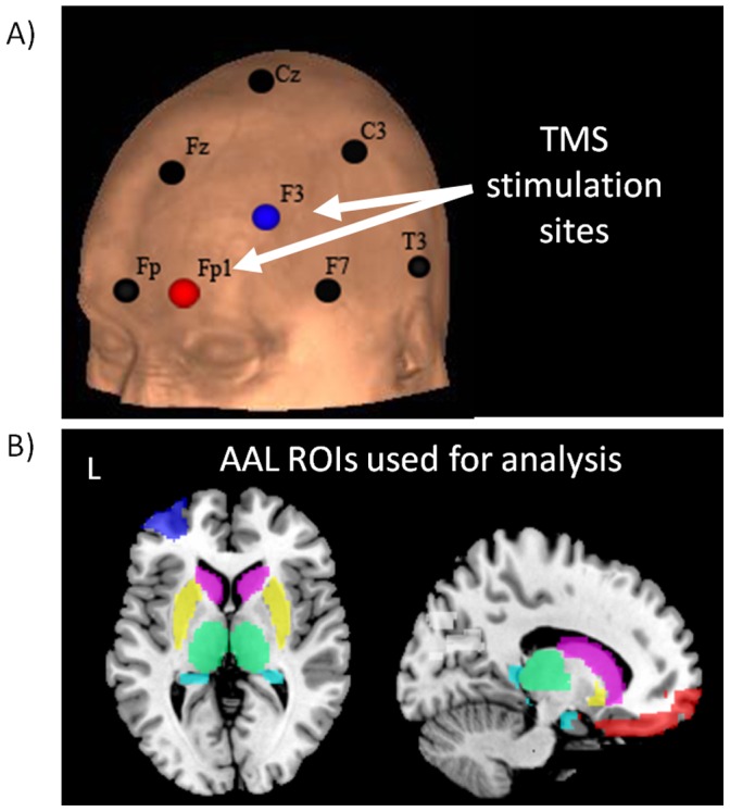
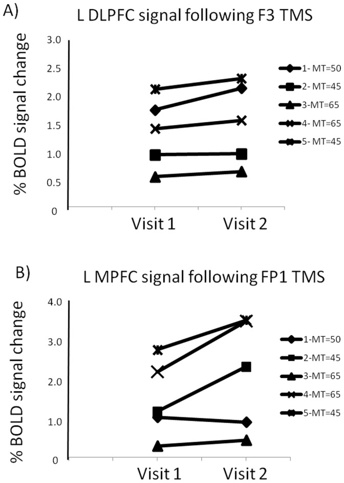
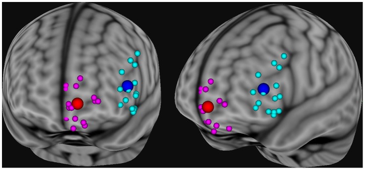
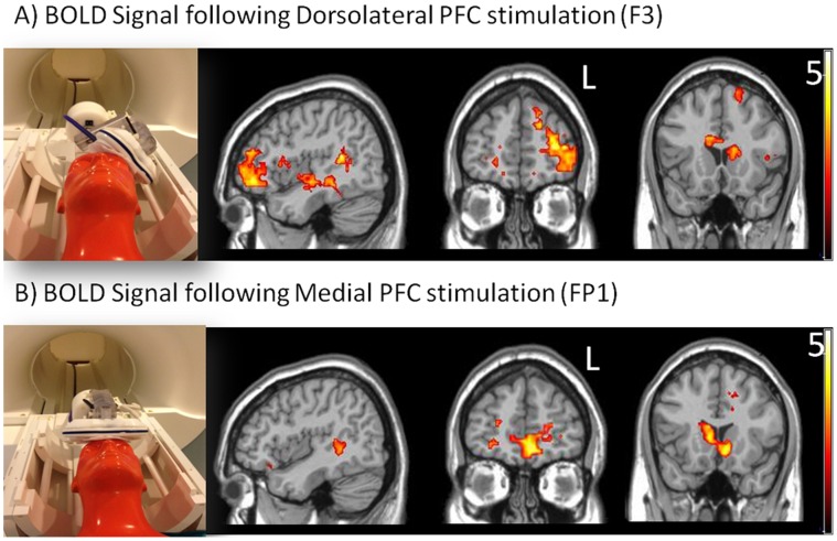
Seventeen healthy individuals received interleaved TMS-BOLD imaging with the coil positioned over the dorsolateral (EEG: F3) and ventromedial PFC (EEG: FP1). BOLD signal change was calculated in the areas directly stimulated by the coil and in subcortical regions with afferent and efferent connectivity to the TMS target areas. Additionally, five individuals were tested on two occasions to determine test-retest reliability.
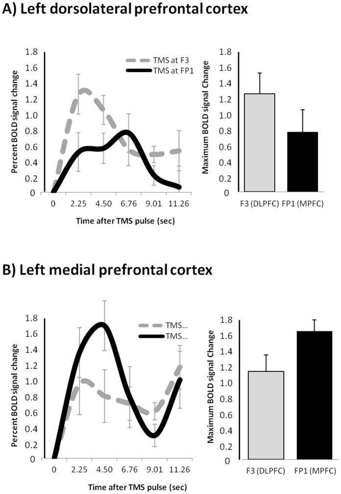
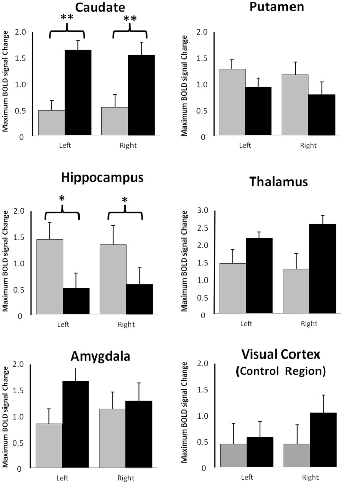
Region of interest analysis revealed that TMS at both prefrontal sites led to significant BOLD signal increases in the cortex under the coil, in the striatum, and the thalamus, but not in the visual cortex (negative control region). There was a significantly larger BOLD signal change in the caudate following medial PFC TMS, relative to lateral TMS. The hippocampus in contrast was significantly more activated by lateral TMS. Post-hoc voxel-based analysis revealed that within the caudate the location of peak activity was in the ventral caudate following medial TMS and the dorsal caudate following lateral TMS. Test-retest reliability data revealed consistent BOLD responses to TMS within each individual but a large variation between individuals.
These data demonstrate that, through an optimized TMS/BOLD sequence over two unique prefrontal targets, it is possible to selectively interrogate the patency of these established cortical-subcortical networks in healthy individuals, and potentially patient populations.
前额叶皮层(PFC)是一个解剖和功能上具有异质性的区域,通过与皮质下靶点的连接影响认知和边缘处理。正如 Alexander 等人(1986 年)所提出的,PFC 的外侧和内侧部分以并行但功能不同的回路投射到纹状体的不同区域。本初步研究的目的是确定是否可以通过在磁共振环境中交错的经颅磁刺激(TMS),对涉及执行和边缘处理的这些外侧和内侧皮质-皮质下回路进行不同且一致的激活。
17 名健康个体接受了线圈置于背外侧(EEG:F3)和腹内侧前额叶皮层(EEG:FP1)上方的交错 TMS-BOLD 成像。通过线圈直接刺激的区域和与 TMS 靶区有传入和传出连接的皮质下区域计算 BOLD 信号变化。此外,有 5 名个体在两次测试中进行了测试,以确定测试-重测可靠性。
感兴趣区分析显示,在前额叶两个部位的 TMS 都导致了线圈下皮层、纹状体和丘脑的 BOLD 信号显著增加,但视觉皮层(阴性对照区)没有。与外侧 TMS 相比,内侧 PFC TMS 后尾状核的 BOLD 信号变化明显更大。相反,外侧 TMS 更能激活海马。基于体素的事后分析显示,在尾状核内,内侧 TMS 后活性峰值位于尾状核腹侧,而外侧 TMS 后位于尾状核背侧。测试-重测可靠性数据显示,每个个体对 TMS 的 BOLD 反应一致,但个体之间的变化很大。
这些数据表明,通过在两个独特的前额叶靶点上优化 TMS/BOLD 序列,可以选择性地检测健康个体中这些已建立的皮质-皮质下网络的通畅性,并可能检测患者群体。