Gossan Nicole, Zeef Leo, Hensman James, Hughes Alun, Bateman John F, Rowley Lynn, Little Christopher B, Piggins Hugh D, Rattray Magnus, Boot-Handford Raymond P, Meng Qing-Jun
University of Manchester, Manchester, UK.
Arthritis Rheum. 2013 Sep;65(9):2334-45. doi: 10.1002/art.38035.
To characterize the circadian clock in murine cartilage tissue and identify tissue-specific clock target genes, and to investigate whether the circadian clock changes during aging or during cartilage degeneration using an experimental mouse model of osteoarthritis (OA).
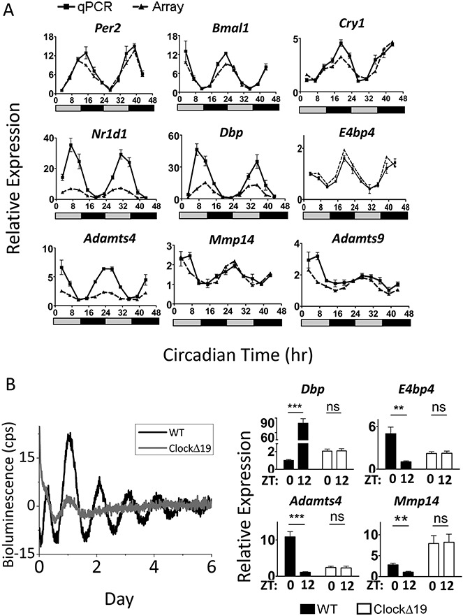
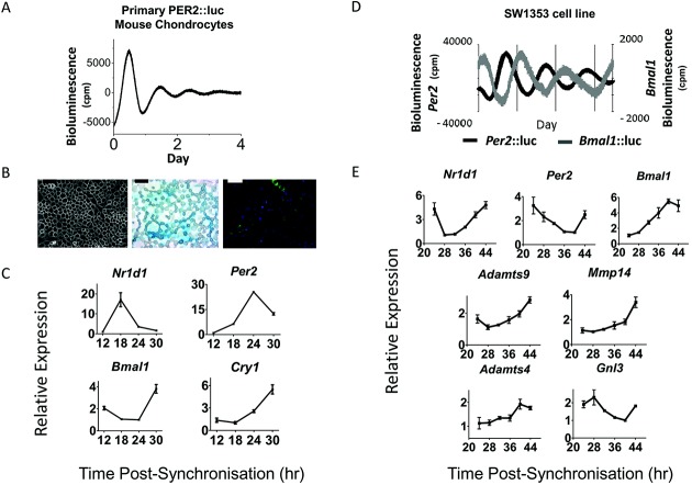
Cartilage explants were obtained from aged and young adult mice after transduction with the circadian clock fusion protein reporter PER2::luc, and real-time bioluminescence recordings were used to characterize the properties of the clock. Time-series microarrays were performed on mouse cartilage tissue to identify genes expressed in a circadian manner. Rhythmic genes were confirmed by quantitative reverse transcription-polymerase chain reaction using mouse tissue, primary chondrocytes, and a human chondrocyte cell line. Experimental OA was induced in mice by destabilization of the medial meniscus (DMM), and articular cartilage samples were microdissected and subjected to microarray analysis.
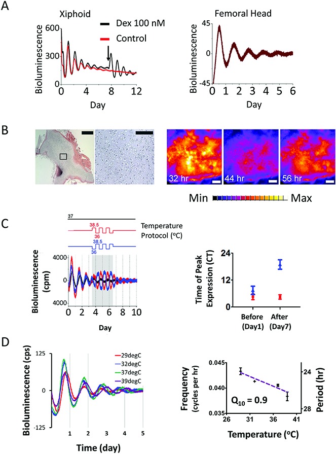
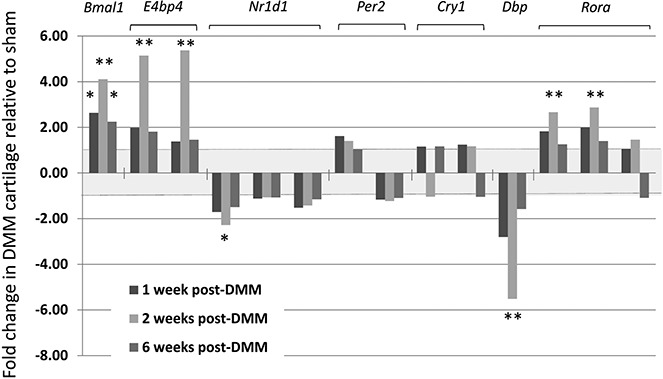
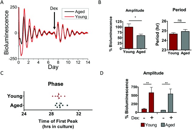
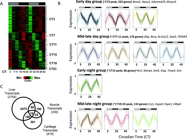
Mouse cartilage tissue and a human chondrocyte cell line were found to contain intrinsic molecular circadian clocks. The cartilage clock could be reset by temperature signals, while the circadian period was temperature compensated. PER2::luc bioluminescence demonstrated that circadian oscillations were significantly lower in amplitude in cartilage from aged mice. Time-series microarray analyses of the mouse tissue identified the first circadian transcriptome in cartilage, revealing that 615 genes (∼3.9% of the expressed genes) displayed a circadian pattern of expression. This included genes involved in cartilage homeostasis and survival, as well as genes with potential importance in the pathogenesis of OA. Several clock genes were disrupted in the early stages of cartilage degeneration in the DMM mouse model of OA.
These results reveal an autonomous circadian clock in chondrocytes that can be implicated in key aspects of cartilage biology and pathology. Consequently, circadian disruption (e.g., during aging) may compromise tissue homeostasis and increase susceptibility to joint damage or disease.
表征小鼠软骨组织中的昼夜节律时钟并鉴定组织特异性时钟靶基因,使用骨关节炎(OA)实验小鼠模型研究昼夜节律时钟在衰老或软骨退变过程中是否发生变化。
在用昼夜节律时钟融合蛋白报告基因PER2::luc转导后,从老年和年轻成年小鼠获取软骨外植体,并使用实时生物发光记录来表征时钟特性。对小鼠软骨组织进行时间序列微阵列分析以鉴定以昼夜节律方式表达的基因。使用小鼠组织、原代软骨细胞和人软骨细胞系通过定量逆转录 - 聚合酶链反应确认节律性基因。通过内侧半月板不稳定(DMM)在小鼠中诱导实验性OA,对关节软骨样本进行显微切割并进行微阵列分析。
发现小鼠软骨组织和人软骨细胞系含有内在分子昼夜节律时钟。软骨时钟可被温度信号重置,而昼夜周期具有温度补偿性。PER2::luc生物发光表明老年小鼠软骨中的昼夜节律振荡幅度显著降低。对小鼠组织的时间序列微阵列分析确定了软骨中的首个昼夜转录组,揭示615个基因(约占表达基因的3.9%)呈现昼夜节律表达模式。这包括参与软骨稳态和存活的基因,以及在OA发病机制中具有潜在重要性的基因。在OA的DMM小鼠模型中,软骨退变早期几个时钟基因被破坏。
这些结果揭示了软骨细胞中存在自主昼夜节律时钟,其可能与软骨生物学和病理学的关键方面有关。因此,昼夜节律紊乱(例如在衰老过程中)可能损害组织稳态并增加对关节损伤或疾病的易感性。