Department of Medicine and Liver Center, Yale University School of Medicine, New Haven, Connecticut, USA.
Compr Physiol. 2013 Jul;3(3):1035-78. doi: 10.1002/cphy.c120027.
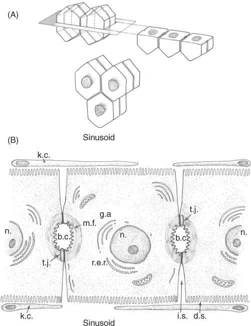
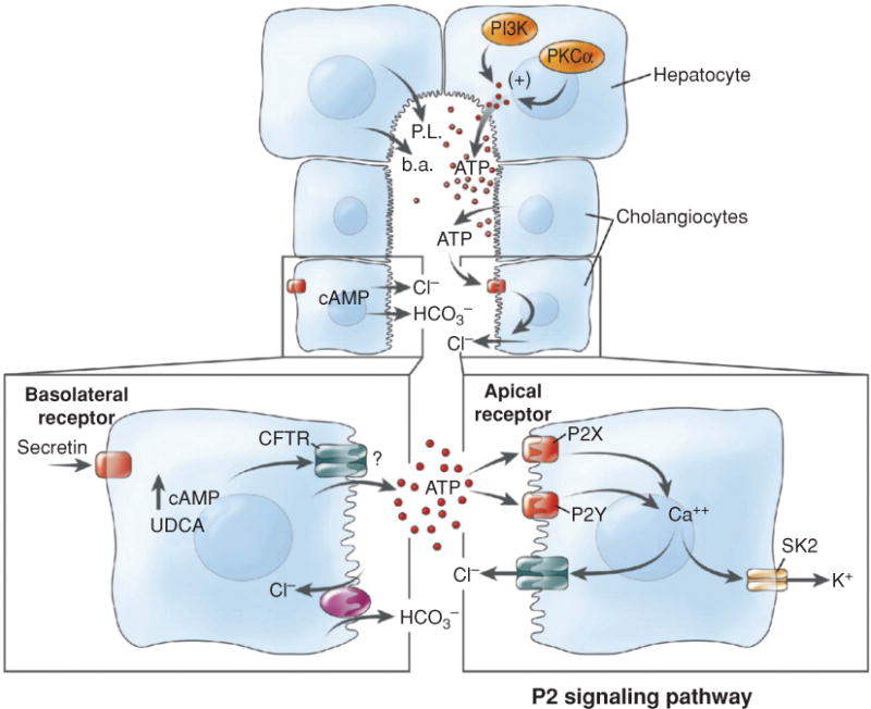
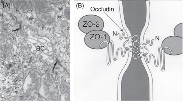
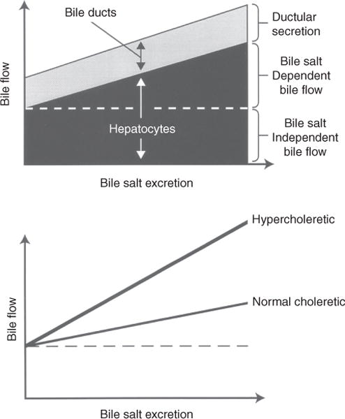
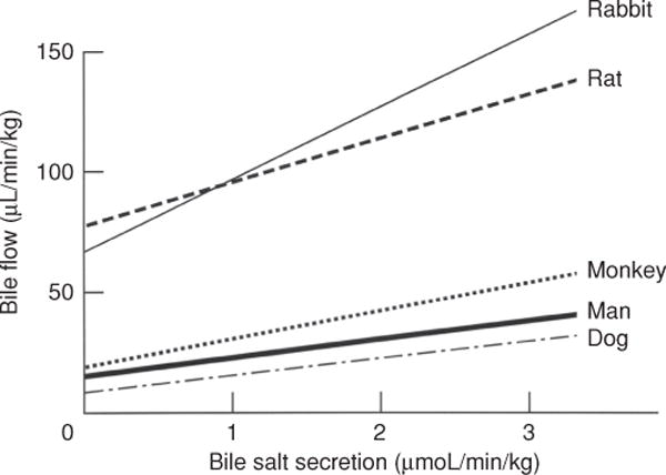
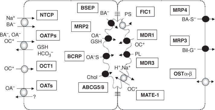
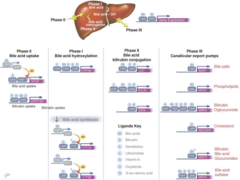
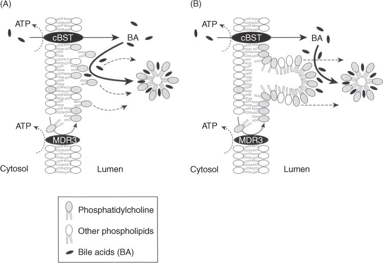
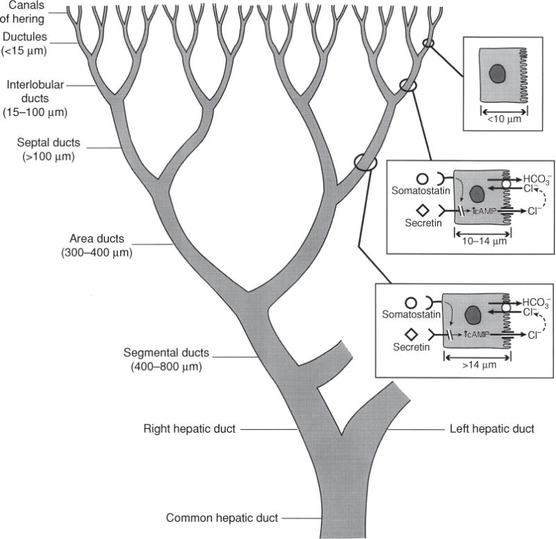
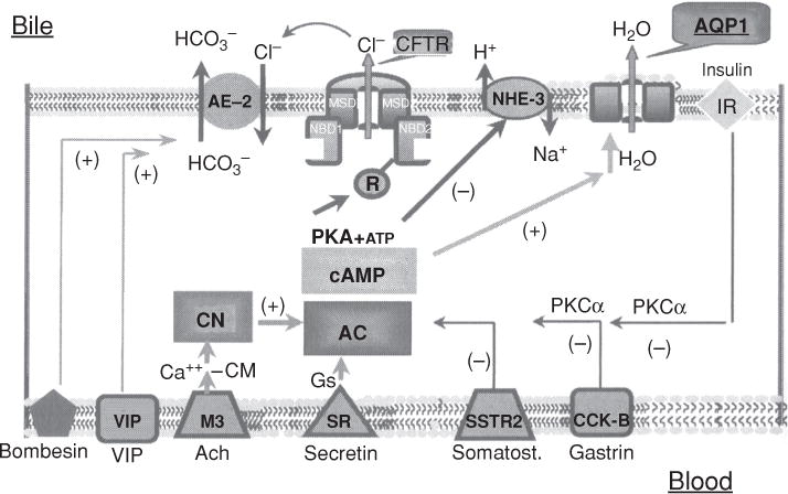
Bile is a unique and vital aqueous secretion of the liver that is formed by the hepatocyte and modified down stream by absorptive and secretory properties of the bile duct epithelium. Approximately 5% of bile consists of organic and inorganic solutes of considerable complexity. The bile-secretory unit consists of a canalicular network which is formed by the apical membrane of adjacent hepatocytes and sealed by tight junctions. The bile canaliculi (∼1 μm in diameter) conduct the flow of bile countercurrent to the direction of portal blood flow and connect with the canal of Hering and bile ducts which progressively increase in diameter and complexity prior to the entry of bile into the gallbladder, common bile duct, and intestine. Canalicular bile secretion is determined by both bile salt-dependent and independent transport systems which are localized at the apical membrane of the hepatocyte and largely consist of a series of adenosine triphosphate-binding cassette transport proteins that function as export pumps for bile salts and other organic solutes. These transporters create osmotic gradients within the bile canalicular lumen that provide the driving force for movement of fluid into the lumen via aquaporins. Species vary with respect to the relative amounts of bile salt-dependent and independent canalicular flow and cholangiocyte secretion which is highly regulated by hormones, second messengers, and signal transduction pathways. Most determinants of bile secretion are now characterized at the molecular level in animal models and in man. Genetic mutations serve to illuminate many of their functions.
胆汁是肝脏分泌的一种独特而重要的水性分泌物,由肝细胞形成,并通过胆管上皮细胞的吸收和分泌特性进行修饰。大约 5%的胆汁由相当复杂的有机和无机溶质组成。胆汁分泌单位由相邻肝细胞的顶膜形成的管腔网络组成,并通过紧密连接密封。胆汁小管(直径约 1μm)引导与门静脉血流方向相反的胆汁流动,并与赫令管和胆管相连,在胆汁进入胆囊、胆总管和肠道之前,胆管逐渐增大并变得更加复杂。胆汁盐依赖和非依赖的转运系统决定了胆汁的分泌,这些系统定位于肝细胞的顶膜,主要由一系列三磷酸腺苷结合盒转运蛋白组成,这些蛋白作为胆汁盐和其他有机溶质的输出泵发挥作用。这些转运蛋白在胆汁小管腔内部创建渗透梯度,为液体通过水通道蛋白进入管腔提供驱动力。不同物种之间胆汁盐依赖和非依赖的胆汁分泌以及胆管细胞分泌的相对量存在差异,这受到激素、第二信使和信号转导途径的高度调节。大多数胆汁分泌的决定因素现在在动物模型和人类中都在分子水平上得到了描述。遗传突变有助于阐明它们的许多功能。