Macfarlane Matthew D, Looi Jeffrey C L, Walterfang Mark, Spulber Gabriela, Velakoulis Dennis, Styner Martin, Crisby Milita, Orndahl Eva, Erkinjuntti Timo, Waldemar Gunhild, Garde Ellen, Hennerici Michael G, Bäzner Hansjörg, Blahak Christian, Wallin Anders, Wahlund Lars-Olof
Research Centre for the Neurosciences of Ageing, Academic Unit of Psychiatry and Addiction Medicine, Australian National University Medical School, Canberra Hospital, Canberra, Australian Capital Territory, Australia.
Research Centre for the Neurosciences of Ageing, Academic Unit of Psychiatry and Addiction Medicine, Australian National University Medical School, Canberra Hospital, Canberra, Australian Capital Territory, Australia; Division of Clinical Geriatrics, Department of Neurobiology, Care Science and Society, Karolinska Institute, Stockholm, Sweden.
Am J Geriatr Psychiatry. 2015 Jan;23(1):59-71.e1. doi: 10.1016/j.jagp.2013.04.011. Epub 2013 Aug 1.
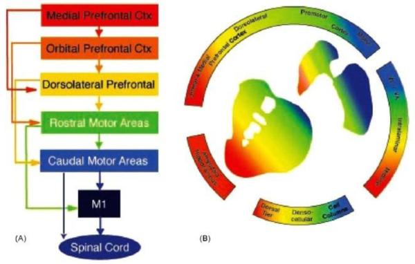
Functional deficits seen in several neurodegenerative disorders have been linked with dysfunction in frontostriatal circuits and with associated shape alterations in striatal structures. The severity of visible white matter hyperintensities (WMHs) on magnetic resonance imaging has been found to correlate with poorer performance on measures of gait and balance. This study aimed to determine whether striatal volume and shape changes were correlated with gait dysfunction.
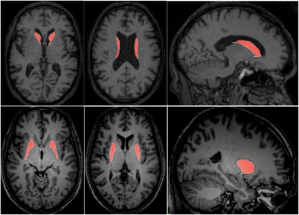
Magnetic resonance imaging scans and clinical gait/balance data (scores from the Short Physical Performance Battery [SPPB]) were sourced from 66 subjects in the previously published LADIS trial, performed in nondisabled individuals older than age 65 years with WMHs at study entry. Data were obtained at study entry and at 3-year follow-up. Caudate nuclei and putamina were manually traced using a previously published method and volumes calculated. The relationships between volume and physical performance on the SPPB were investigated with shape analysis using the spherical harmonic shape description toolkit.
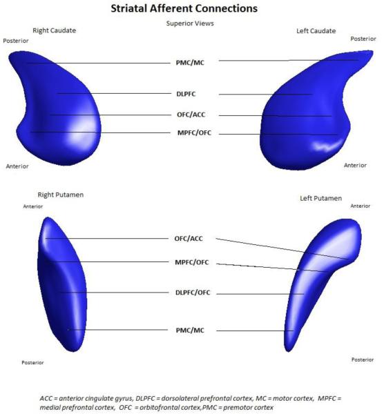
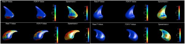
There was no correlation between the severity of WMHs and striatal volumes. Caudate nuclei volume correlated with performance on the SPPB at baseline but not at follow-up, with subsequent shape analysis showing left caudate changes occurred in areas corresponding to inputs of the dorsolateral prefrontal, premotor, and motor cortex. There was no correlation between putamen volumes and performance on the SPPB.
Disruption in frontostriatal circuits may play a role in mediating poorer physical performance in individuals with WMHs. Striatal volume and shape changes may be suitable biomarkers for functional changes in this population.
在几种神经退行性疾病中观察到的功能缺陷与额纹状体回路功能障碍以及纹状体结构的相关形态改变有关。磁共振成像中可见的白质高信号(WMHs)的严重程度已被发现与步态和平衡测量中的较差表现相关。本研究旨在确定纹状体体积和形状变化是否与步态功能障碍相关。
磁共振成像扫描和临床步态/平衡数据(简短身体功能测试[SPPB]的分数)来自先前发表的LADIS试验中的66名受试者,该试验在65岁以上、入组时患有WMHs的非残疾个体中进行。在入组时和3年随访时获取数据。使用先前发表的方法手动描绘尾状核和壳核,并计算体积。使用球谐形状描述工具包通过形状分析研究体积与SPPB上身体表现之间的关系。
WMHs的严重程度与纹状体体积之间没有相关性。尾状核体积在基线时与SPPB上的表现相关,但在随访时不相关,随后的形状分析表明左侧尾状核的变化发生在与背外侧前额叶、运动前区和运动皮层输入相对应的区域。壳核体积与SPPB上的表现之间没有相关性。
额纹状体回路的破坏可能在介导患有WMHs的个体身体表现较差方面起作用。纹状体体积和形状变化可能是该人群功能变化的合适生物标志物。