Turner Lauren M, Jakabek David, Wilkes Fiona A, Croft Rodney J, Churchyard Andrew, Walterfang Mark, Velakoulis Dennis, Looi Jeffrey C L, Georgiou-Karistianis Nellie, Apthorp Deborah
Research School of Psychology College of Medicine, Biology, & Environment Australian National University Canberra Australian Capital Territory Australia.
Graduate School of Medicine University of Wollongong Wollongong New South Wales Australia.
Brain Behav. 2016 Jul 27;6(12):e00511. doi: 10.1002/brb3.511. eCollection 2016 Dec.
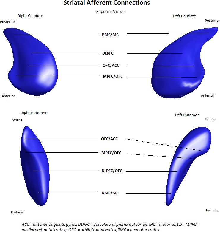
Huntington's disease (HD) causes progressive atrophy to the striatum, a critical node in frontostriatal circuitry. Maintenance of motor function is dependent on functional connectivity of these premotor, motor, and dorsolateral frontostriatal circuits, and structural integrity of the striatum itself. We aimed to investigate whether size and shape of the striatum as a measure of frontostriatal circuit structural integrity was correlated with functional frontostriatal electrophysiological neural premotor processing (contingent negative variation, CNV), to better understand motoric structure-function relationships in early HD.
Magnetic resonance imaging (MRI) scans and electrophysiological (EEG) measures of premotor processing were obtained from a combined HD group (12 presymptomatic, 7 symptomatic). Manual segmentation of caudate and putamen was conducted with subsequent shape analysis. Separate correlational analyses (volume and shape) included covariates of age, gender, intracranial volume, and time between EEG and MRI.
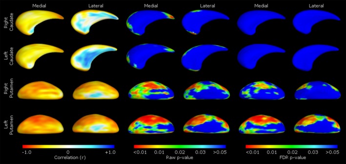
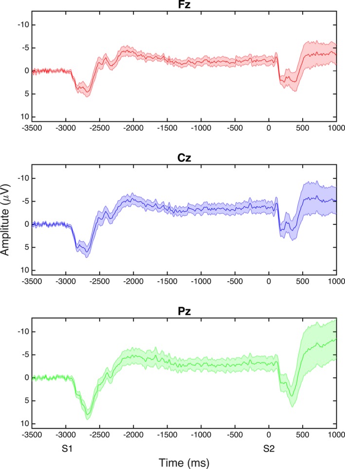
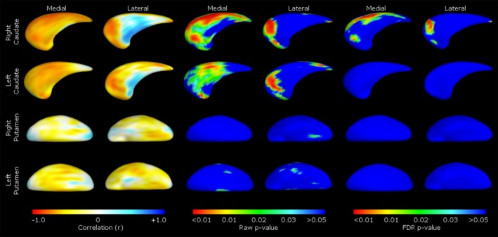
Right caudate volume correlated with early CNV latency over frontocentral regions and late CNV frontally, whereas right caudate shape correlated with early CNV latency centrally. Left caudate volume correlated with early CNV latency over centroparietal regions and late CNV frontally. Right and left putamen volumes correlated with early CNV latency frontally, and right and left putamen shape/volume correlated with parietal CNV slope.
Timing (latency) and pattern (slope) of frontostriatal circuit-mediated premotor functional activation across scalp regions were correlated with abnormalities in structural integrity of the key frontostriatal circuit component, the striatum (size and shape). This was accompanied by normal reaction times, suggesting it may be undetected in regular tasks due to preserved motor "performance." Such differences in functional activation may reflect atrophy-based frontostriatal circuitry despecialization and/or compensatory recruitment of additional brain regions.
亨廷顿舞蹈症(HD)会导致纹状体进行性萎缩,而纹状体是额纹状体回路中的关键节点。运动功能的维持依赖于这些运动前区、运动区和背外侧额纹状体回路的功能连接,以及纹状体本身的结构完整性。我们旨在研究作为额纹状体回路结构完整性指标的纹状体大小和形状是否与额纹状体电生理神经运动前处理(关联性负变,CNV)相关,以更好地理解早期HD中的运动结构-功能关系。
从一个合并的HD组(12名症状前患者,7名有症状患者)获取磁共振成像(MRI)扫描和运动前处理的电生理(EEG)测量数据,并对尾状核和壳核进行手动分割及后续形状分析。单独的相关性分析(体积和形状)纳入了年龄、性别、颅内体积以及EEG和MRI之间时间间隔等协变量。
右侧尾状核体积与额中央区早期CNV潜伏期以及额叶晚期CNV相关,而右侧尾状核形状与中央区早期CNV潜伏期相关。左侧尾状核体积与顶中央区早期CNV潜伏期以及额叶晚期CNV相关。右侧和左侧壳核体积与额叶早期CNV潜伏期相关,右侧和左侧壳核形状/体积与顶叶CNV斜率相关。
额纹状体回路介导的头皮区域运动前功能激活的时间(潜伏期)和模式(斜率)与关键额纹状体回路组成部分纹状体(大小和形状)的结构完整性异常相关。这伴随着正常的反应时间,表明由于运动“表现”得以保留,在常规任务中可能无法检测到。这种功能激活的差异可能反映了基于萎缩的额纹状体回路去特化和/或额外脑区的代偿性募集。