• 文献检索
  • 文档翻译
  • 深度研究
  • 学术资讯
  • Suppr Zotero 插件Zotero 插件
  • 邀请有礼
  • 套餐&价格
  • 历史记录
应用&插件
Suppr Zotero 插件Zotero 插件浏览器插件Mac 客户端Windows 客户端微信小程序
定价
高级版会员购买积分包购买API积分包
服务
文献检索文档翻译深度研究API 文档MCP 服务
关于我们
关于 Suppr公司介绍联系我们用户协议隐私条款
关注我们

Suppr 超能文献

核心技术专利:CN118964589B侵权必究
粤ICP备2023148730 号-1Suppr @ 2026

文献检索

告别复杂PubMed语法,用中文像聊天一样搜索,搜遍4000万医学文献。AI智能推荐,让科研检索更轻松。

立即免费搜索

文件翻译

保留排版,准确专业,支持PDF/Word/PPT等文件格式,支持 12+语言互译。

免费翻译文档

深度研究

AI帮你快速写综述,25分钟生成高质量综述,智能提取关键信息,辅助科研写作。

立即免费体验

3D 纹理分析显示进行性肌阵挛性癫痫 1 型(EPM1)患者丘脑和壳核的 MRI 纹理改变难以察觉。

3D texture analysis reveals imperceptible MRI textural alterations in the thalamus and putamen in progressive myoclonic epilepsy type 1, EPM1.

机构信息

Department of Clinical Radiology, Kuopio University Hospital, Kuopio, Finland.

出版信息

PLoS One. 2013 Jul 29;8(7):e69905. doi: 10.1371/journal.pone.0069905. Print 2013.

DOI:10.1371/journal.pone.0069905
PMID:23922849
原文链接:https://pmc.ncbi.nlm.nih.gov/articles/PMC3726751/
Abstract

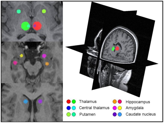
Progressive myoclonic epilepsy type 1 (EPM1) is an autosomal recessively inherited neurodegenerative disorder characterized by young onset age, myoclonus and tonic-clonic epileptic seizures. At the time of diagnosis, the visual assessment of the brain MRI is usually normal, with no major changes found later. Therefore, we utilized texture analysis (TA) to characterize and classify the underlying properties of the affected brain tissue by means of 3D texture features. Sixteen genetically verified patients with EPM1 and 16 healthy controls were included in the study. TA was performed upon 3D volumes of interest that were placed bilaterally in the thalamus, amygdala, hippocampus, caudate nucleus and putamen. Compared to the healthy controls, EPM1 patients had significant textural differences especially in the thalamus and right putamen. The most significantly differing texture features included parameters that measure the complexity and heterogeneity of the tissue, such as the co-occurrence matrix-based entropy and angular second moment, and also the run-length matrix-based parameters of gray-level non-uniformity, short run emphasis and long run emphasis. This study demonstrates the usability of 3D TA for extracting additional information from MR images. Textural alterations which suggest complex, coarse and heterogeneous appearance were found bilaterally in the thalamus, supporting the previous literature on thalamic pathology in EPM1. The observed putamenal involvement is a novel finding. Our results encourage further studies on the clinical applications, feasibility, reproducibility and reliability of 3D TA.

摘要

进行性肌阵挛癫痫 1 型(EPM1)是一种常染色体隐性遗传性神经退行性疾病,其特征为发病年龄早、肌阵挛和强直阵挛性癫痫发作。在诊断时,脑 MRI 的视觉评估通常正常,随后也没有发现重大变化。因此,我们利用纹理分析(TA)通过 3D 纹理特征来描述和分类受影响脑组织的潜在特性。本研究纳入了 16 名经基因证实的 EPM1 患者和 16 名健康对照者。TA 是在双侧丘脑、杏仁核、海马体、尾状核和壳核的 3D 感兴趣区容积上进行的。与健康对照组相比,EPM1 患者的纹理存在显著差异,尤其是在丘脑和右侧壳核。差异最显著的纹理特征包括测量组织复杂性和异质性的参数,如基于共生矩阵的熵和角二阶矩,以及基于游程长度矩阵的灰度不均匀性、短游程强调和长游程强调的参数。本研究证明了 3D TA 从 MR 图像中提取额外信息的可用性。在丘脑双侧发现了纹理改变,提示存在复杂、粗糙和异质性外观,这支持了先前关于 EPM1 中丘脑病理学的文献。观察到壳核受累是一个新发现。我们的研究结果鼓励进一步研究 3D TA 的临床应用、可行性、可重复性和可靠性。

https://cdn.ncbi.nlm.nih.gov/pmc/blobs/a4ae/3726751/34a0fcb43331/pone.0069905.g002.jpg
https://cdn.ncbi.nlm.nih.gov/pmc/blobs/a4ae/3726751/42c165aa2e67/pone.0069905.g001.jpg
https://cdn.ncbi.nlm.nih.gov/pmc/blobs/a4ae/3726751/34a0fcb43331/pone.0069905.g002.jpg
https://cdn.ncbi.nlm.nih.gov/pmc/blobs/a4ae/3726751/42c165aa2e67/pone.0069905.g001.jpg
https://cdn.ncbi.nlm.nih.gov/pmc/blobs/a4ae/3726751/34a0fcb43331/pone.0069905.g002.jpg

相似文献

1
3D texture analysis reveals imperceptible MRI textural alterations in the thalamus and putamen in progressive myoclonic epilepsy type 1, EPM1.3D 纹理分析显示进行性肌阵挛性癫痫 1 型(EPM1)患者丘脑和壳核的 MRI 纹理改变难以察觉。
PLoS One. 2013 Jul 29;8(7):e69905. doi: 10.1371/journal.pone.0069905. Print 2013.
2
Progressive myoclonic epilepsy type 1 (EPM1) patients present with abnormal H MRS brain metabolic profiles associated with cognitive function.进行性肌阵挛癫痫 1 型(EPM1)患者存在异常的 H MRS 脑代谢谱,与认知功能有关。
Neuroimage Clin. 2023;39:103459. doi: 10.1016/j.nicl.2023.103459. Epub 2023 Jul 3.
3
Subcortical gray matter changes in pediatric patients with new-onset juvenile myoclonic epilepsy.儿童新诊断的青少年肌阵挛性癫痫患者的皮质下灰质变化。
Epilepsy Behav. 2020 Mar;104(Pt A):106860. doi: 10.1016/j.yebeh.2019.106860. Epub 2020 Jan 11.
4
Developmentally stable whole-brain volume reductions and developmentally sensitive caudate and putamen volume alterations in those with attention-deficit/hyperactivity disorder and their unaffected siblings.患有注意缺陷多动障碍(ADHD)及其未受影响的兄弟姐妹存在全脑体积发育稳定减少和尾状核和壳核体积发育敏感改变。
JAMA Psychiatry. 2015 May;72(5):490-9. doi: 10.1001/jamapsychiatry.2014.3162.
5
Children with epilepsy demonstrate macro- and microstructural changes in the thalamus, putamen, and amygdala.患有癫痫的儿童的丘脑、壳核和杏仁核存在宏观和微观结构的变化。
Neuroradiology. 2020 Mar;62(3):389-397. doi: 10.1007/s00234-019-02332-8. Epub 2019 Dec 18.
6
Patients with extratemporal lobe epilepsy do not differ from healthy subjects with respect to subcortical volumes.颞叶外癫痫患者在皮质下体积方面与健康受试者并无差异。
J Neurol Neurosurg Psychiatry. 2004 Apr;75(4):588-92. doi: 10.1136/jnnp.2003.018721.
7
Strongly reduced volumes of putamen and thalamus in Alzheimer's disease: an MRI study.阿尔茨海默病中壳核和丘脑体积显著减小:一项MRI研究。
Brain. 2008 Dec;131(Pt 12):3277-85. doi: 10.1093/brain/awn278. Epub 2008 Nov 20.
8
Neurobehavioral comorbidities of pediatric epilepsies are linked to thalamic structural abnormalities.儿科癫痫的神经行为共病与丘脑结构异常有关。
Epilepsia. 2013 Dec;54(12):2116-24. doi: 10.1111/epi.12428. Epub 2013 Nov 8.
9
Subcortical structural abnormalities in juvenile myoclonic epilepsy (JME): MR volumetry and vertex based analysis.青少年肌阵挛癫痫(JME)的皮质下结构异常:MR 容积测量和基于顶点的分析。
Seizure. 2013 Apr;22(3):230-5. doi: 10.1016/j.seizure.2013.01.001. Epub 2013 Jan 20.
10
Motor cortex and thalamic atrophy in Unverricht-Lundborg disease: voxel-based morphometric study.乌-伦二氏病中运动皮层和丘脑萎缩:基于体素的形态学研究
Neurology. 2009 Aug 25;73(8):606-11. doi: 10.1212/WNL.0b013e3181b3888b.

引用本文的文献

1
Molecular classification of patients with grade II/III glioma using quantitative MRI characteristics.基于定量 MRI 特征的 II/III 级脑胶质瘤患者的分子分类。
J Neurooncol. 2018 Sep;139(3):633-642. doi: 10.1007/s11060-018-2908-3. Epub 2018 Jun 2.
2
Edge Contrast of the FLAIR Hyperintense Region Predicts Survival in Patients with High-Grade Gliomas following Treatment with Bevacizumab.FLAIR 高信号区域的边缘对比度预测贝伐珠单抗治疗后高级别胶质瘤患者的生存。
AJNR Am J Neuroradiol. 2018 Jun;39(6):1017-1024. doi: 10.3174/ajnr.A5620. Epub 2018 Apr 5.
3
Imaging of Tau Pathology Using Magnetic Resonance Imaging Textural Analysis.

本文引用的文献

1
Texture analysis of magnetic resonance images of patients with juvenile myoclonic epilepsy.患者磁共振图像纹理分析在青少年肌阵挛性癫痫中的应用。
Epilepsy Behav. 2013 Apr;27(1):22-8. doi: 10.1016/j.yebeh.2012.12.009. Epub 2013 Jan 28.
2
Texture analysis of high resolution MRI allows discrimination between febrile and afebrile initial precipitating injury in mesial temporal sclerosis.高分辨率磁共振成像的纹理分析可区分局灶性内侧颞叶硬化的发热性和无热性初始前驱损伤。
Magn Reson Med. 2012 Nov;68(5):1647-53. doi: 10.1002/mrm.24174. Epub 2012 Jan 27.
3
Sensorimotor, visual, and auditory cortical atrophy in Unverricht-Lundborg disease mapped with cortical thickness analysis.
使用磁共振成像纹理分析对Tau病理进行成像
Front Neurosci. 2017 Nov 6;11:599. doi: 10.3389/fnins.2017.00599. eCollection 2017.
4
2D and 3D texture analysis to differentiate brain metastases on MR images: proceed with caution.二维和三维纹理分析用于在磁共振图像上鉴别脑转移瘤:需谨慎进行。
MAGMA. 2018 Apr;31(2):285-294. doi: 10.1007/s10334-017-0653-9. Epub 2017 Sep 22.
5
Effect of radiation dose reduction on texture measures of trabecular bone microstructure: an in vitro study.辐射剂量降低对小梁骨微观结构纹理测量的影响:一项体外研究。
J Bone Miner Metab. 2018 May;36(3):323-335. doi: 10.1007/s00774-017-0836-5. Epub 2017 Apr 7.
6
Radiomics: a new application from established techniques.放射组学:既定技术的新应用。
Expert Rev Precis Med Drug Dev. 2016;1(2):207-226. doi: 10.1080/23808993.2016.1164013. Epub 2016 Mar 31.
7
MRI texture analysis of subchondral bone at the tibial plateau.胫骨平台软骨下骨的MRI纹理分析
Eur Radiol. 2016 Sep;26(9):3034-45. doi: 10.1007/s00330-015-4142-0. Epub 2015 Dec 17.
8
Hybrid RGSA and Support Vector Machine Framework for Three-Dimensional Magnetic Resonance Brain Tumor Classification.用于三维磁共振脑肿瘤分类的混合RGSA与支持向量机框架
ScientificWorldJournal. 2015;2015:184350. doi: 10.1155/2015/184350. Epub 2015 Oct 4.
皮质厚度分析显示,Unverricht-Lundborg 病患者存在感觉运动、视觉和听觉皮质萎缩。
AJNR Am J Neuroradiol. 2012 May;33(5):878-83. doi: 10.3174/ajnr.A2882. Epub 2012 Jan 19.
4
Early microglial activation precedes neuronal loss in the brain of the Cstb-/- mouse model of progressive myoclonus epilepsy, EPM1.早发性小胶质细胞激活先于进行性肌阵挛癫痫 EPM1 的 Cstb-/- 小鼠模型的大脑神经元丢失。
J Neuropathol Exp Neurol. 2012 Jan;71(1):40-53. doi: 10.1097/NEN.0b013e31823e68e1.
5
3D texture analysis on MRI images of Alzheimer's disease.MRI 图像阿尔茨海默病的三维纹理分析。
Brain Imaging Behav. 2012 Mar;6(1):61-9. doi: 10.1007/s11682-011-9142-3.
6
3D image texture analysis of simulated and real-world vascular trees.模拟和真实血管树的 3D 图像纹理分析。
Comput Methods Programs Biomed. 2012 Aug;107(2):140-54. doi: 10.1016/j.cmpb.2011.06.004. Epub 2011 Jul 30.
7
Microstructural and volumetric abnormalities of the putamen in juvenile myoclonic epilepsy.青少年肌阵挛癫痫患者壳核的微观结构和体积异常。
Epilepsia. 2011 Sep;52(9):1715-24. doi: 10.1111/j.1528-1167.2011.03117.x. Epub 2011 Jun 2.
8
MRI-texture analysis of corpus callosum, thalamus, putamen, and caudate in Machado-Joseph disease.磁共振脑实质纹理分析在脑腱黄瘤病中的应用
J Neuroimaging. 2012 Jan;22(1):46-52. doi: 10.1111/j.1552-6569.2010.00553.x. Epub 2010 Dec 1.
9
Basal ganglia contributions to motor control: a vigorous tutor.基底神经节对运动控制的贡献:一个强有力的指导者。
Curr Opin Neurobiol. 2010 Dec;20(6):704-16. doi: 10.1016/j.conb.2010.08.022. Epub 2010 Sep 17.
10
Mild traumatic brain injury: tissue texture analysis correlated to neuropsychological and DTI findings.轻度创伤性脑损伤:组织纹理分析与神经心理学和 DTI 发现的相关性。
Acad Radiol. 2010 Sep;17(9):1096-102. doi: 10.1016/j.acra.2010.04.009. Epub 2010 Jun 3.