Southern Arizona VA Healthcare System, University of Arizona, Tucson, Arizona, USA.
Indian J Ophthalmol. 2013 Aug;61(8):401-6. doi: 10.4103/0301-4738.116059.
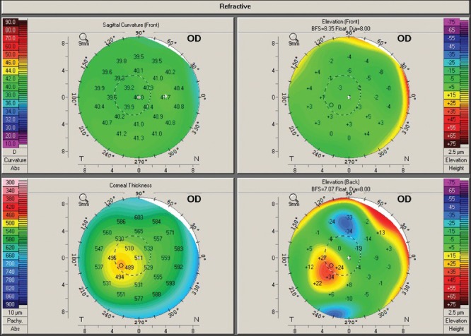
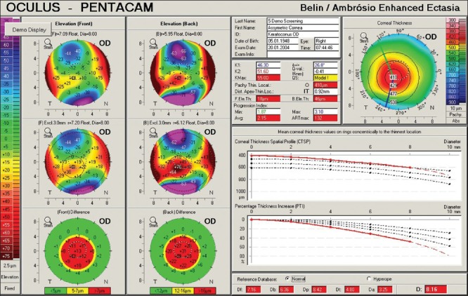
Scheimpflug cross-sectioning anterior segment imaging offers significant advantages over traditional placido based curvature analysis and ultrasound pachymetry. The accurate measurement of both the anterior and posterior corneal surfaces and the anterior and posterior lens allows for the creation of a three-dimensional reconstruction of the anterior segment. Changes on both the posterior cornea and/or corneal thickness map are earlier indicators of ectatic change than would otherwise be identifiable with only anterior curvature and ultrasonic pachymetry. Scheimpflug imaging also covers significantly more of the cornea than was possible with placido based devices. This added coverage is critical in the proper diagnosis of peripheral diseases such as pellucid marginal degeneration (PMD).
Scheimpflug 前节横断面成像相对于传统的基于 Placido 的曲率分析和超声角膜测厚术具有显著优势。对前、后角膜表面以及前、后晶状体的精确测量,可创建前节的三维重建。与仅用前曲率和超声角膜测厚术相比,后角膜和/或角膜厚度图的变化是扩张性病变的早期指标。Scheimpflug 成像也比基于 Placido 的设备能覆盖更多的角膜。这种额外的覆盖范围对于正确诊断周边疾病(如透明边缘变性(PMD))至关重要。