National Institute of Pathology, ICMR, Safdarjung Hospital Campus, New Delhi, India.
PLoS Negl Trop Dis. 2013 Jul 25;7(7):e2338. doi: 10.1371/journal.pntd.0002338. Print 2013.
Patients with localized tuberculoid and generalized lepromatous leprosy show respectively Th1 and Th2 cytokine profile. Additionally, other patients in both types of leprosy also show a non discriminating Th0 cytokine profile with both interferon-γ and IL-4. The present study investigated the role of Th17 cells which appear to be a distinct subtype of Th subtypes in 19 tuberculoid and 18 lepromatous leprosy patients. Five healthy subjects with long term exposure to infection and 4 skin biopsies from healthy subjects undergoing cosmetic surgery were used as controls.
METHODOLOGY/PRINCIPLE FINDINGS: An array of Th17 related primers for cytokines, chemokines and transcription factors was used in real time reverse transcribed PCR to evaluate gene expression, ELISA for cytokine secretion in the supernatants of antigen stimulated PBMC cultures and flow cytometry for establishing the phenotype of the IL-17, IL-21 producing cells.
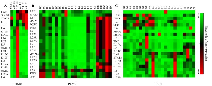
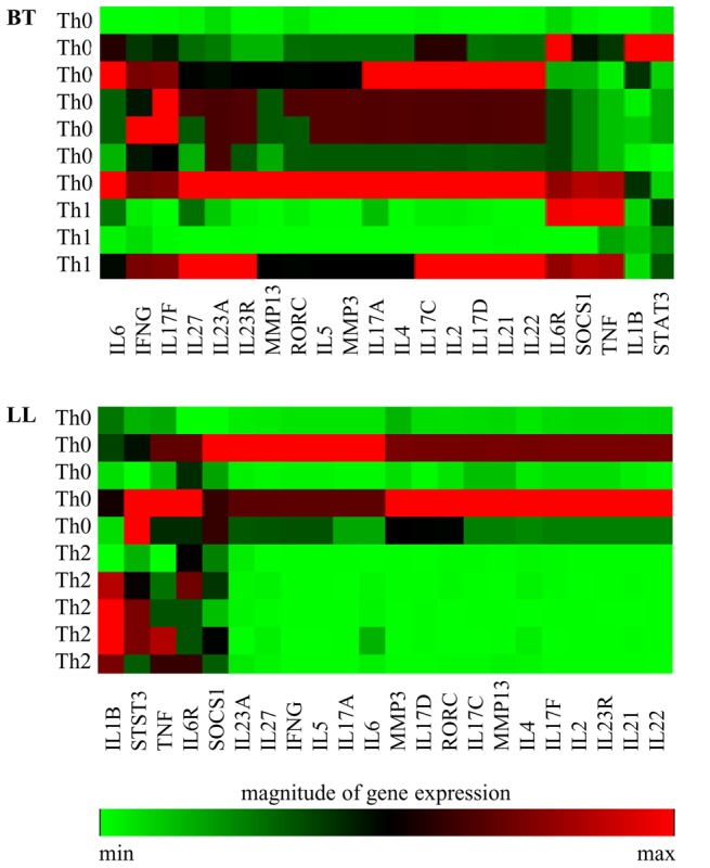
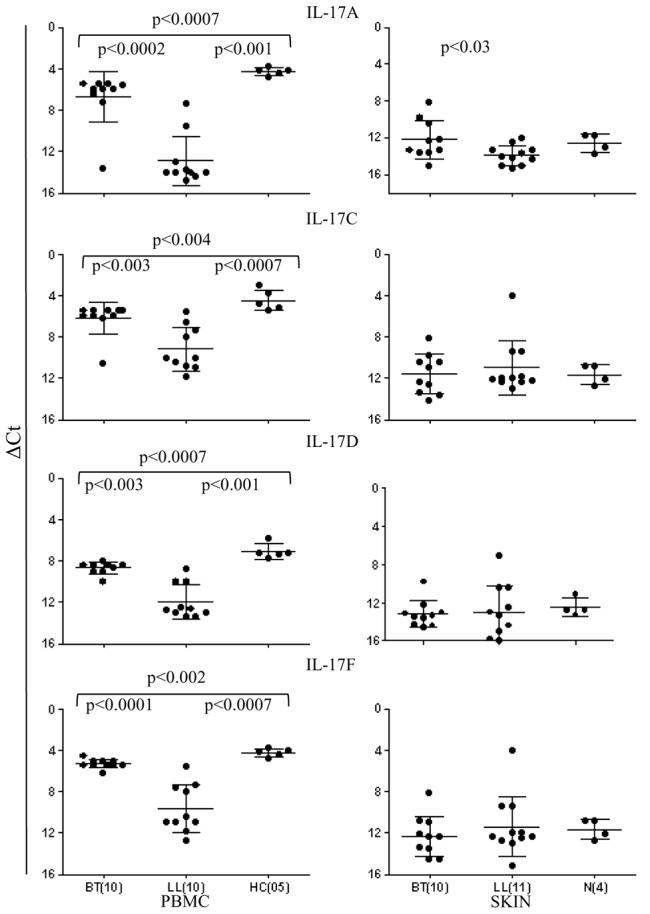
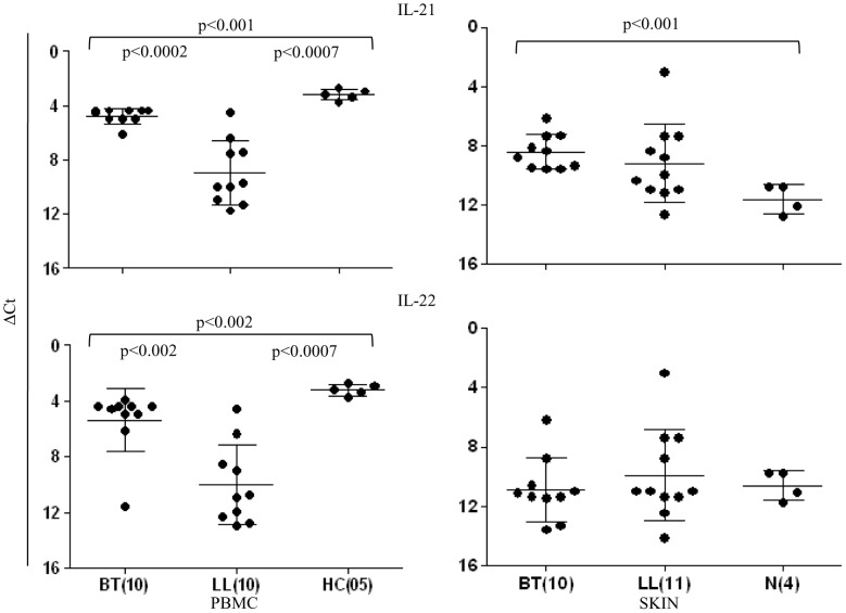
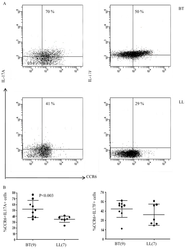
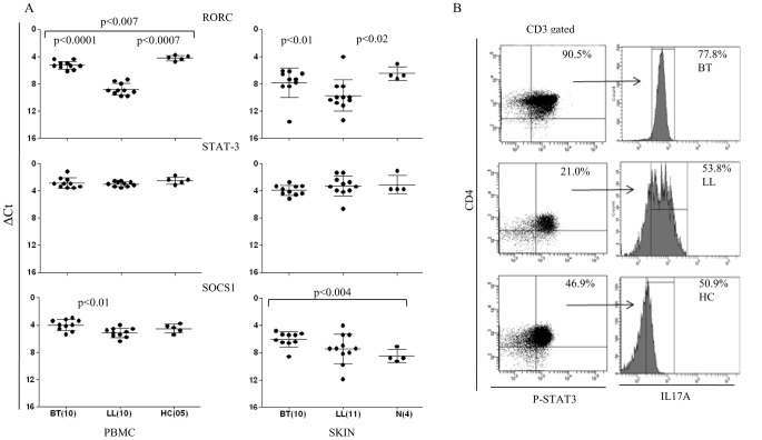
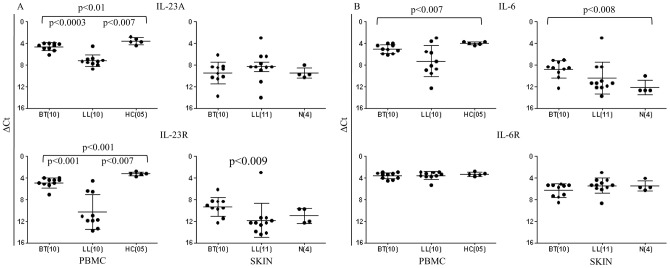
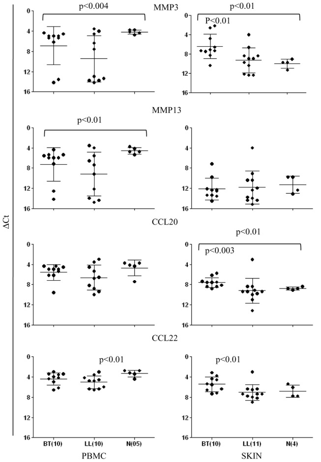
CONCLUSIONS/SIGNIFICANCE: IL-17 isoforms showed significantly higher expression and release in supernatants of antigen stimulated PBMC cultures and dermal lesions of healthy contacts and tuberculoid leprosy as compared to lepromatous leprosy (p<0.003). This was further confirmed by Th17 associated transcription factor RORC, cytokines IL-21, IL-22, and IL-23, chemokines MMP13, CCL20, CCL22. Of interest was the association of IL-23R and not IL-6R with IL-17(+) cells. The Th17 cells were CD4(+) CCR6(+) confirming their effector cell lineage. Polarized Th1 cytokines were seen in 3/7 tuberculoid and Th2 cytokines in 5/10 lepromatous leprosy patients. Of importance was the higher association of Th17 pathway factors with the non-polarized Th0 types as compared to the polarized Th1 and Th2 (p<0.01). Our study draws attention to a third type of effector Th cell that may play a role in leprosy.
局部结核样型和广泛界限类麻风患者分别表现出 Th1 和 Th2 细胞因子谱。此外,两种类型的麻风患者中还有其他患者表现出非特异性 Th0 细胞因子谱,同时存在干扰素-γ和 IL-4。本研究调查了 Th17 细胞在 19 例结核样型和 18 例界限类麻风患者中的作用。5 例长期感染的健康受试者和 4 例接受美容手术的健康受试者的皮肤活检作为对照。
方法/原理发现:使用 Th17 相关细胞因子、趋化因子和转录因子的实时逆转录 PCR 引物阵列评估基因表达,酶联免疫吸附试验(ELISA)检测抗原刺激 PBMC 培养上清液中的细胞因子分泌,流式细胞术分析 IL-17、IL-21 产生细胞的表型。
结论/意义:与界限类麻风相比,抗原刺激 PBMC 培养上清液和健康接触者及结核样型麻风的皮肤病变中 IL-17 同工型的表达和释放显著升高(p<0.003)。这进一步得到了 Th17 相关转录因子 RORC、细胞因子 IL-21、IL-22 和 IL-23、趋化因子 MMP13、CCL20 和 CCL22 的证实。有趣的是,与 IL-17(+)细胞相关的是 IL-23R 而不是 IL-6R。Th17 细胞为 CD4(+)CCR6(+),证实了其效应细胞谱系。3/7 例结核样型患者出现极化 Th1 细胞因子,5/10 例界限类麻风患者出现 Th2 细胞因子。重要的是,与极化 Th1 和 Th2 相比,非极化 Th0 类型与 Th17 途径因子的相关性更高(p<0.01)。我们的研究引起了对可能在麻风病中发挥作用的第三种效应 Th 细胞的关注。