• 文献检索
  • 文档翻译
  • 深度研究
  • 学术资讯
  • Suppr Zotero 插件Zotero 插件
  • 邀请有礼
  • 套餐&价格
  • 历史记录
应用&插件
Suppr Zotero 插件Zotero 插件浏览器插件Mac 客户端Windows 客户端微信小程序
定价
高级版会员购买积分包购买API积分包
服务
文献检索文档翻译深度研究API 文档MCP 服务
关于我们
关于 Suppr公司介绍联系我们用户协议隐私条款
关注我们

Suppr 超能文献

核心技术专利:CN118964589B侵权必究
粤ICP备2023148730 号-1Suppr @ 2026

文献检索

告别复杂PubMed语法,用中文像聊天一样搜索,搜遍4000万医学文献。AI智能推荐,让科研检索更轻松。

立即免费搜索

文件翻译

保留排版,准确专业,支持PDF/Word/PPT等文件格式,支持 12+语言互译。

免费翻译文档

深度研究

AI帮你快速写综述,25分钟生成高质量综述,智能提取关键信息,辅助科研写作。

立即免费体验

腹部放疗中 SPECT/CT 成像的精确肾脏评估(RAPRASI):一项前瞻性试验的设计和方法,旨在提高对肾脏放射剂量反应的理解。

Radiotherapy of abdomen with precise renal assessment with SPECT/CT imaging (RAPRASI): design and methodology of a prospective trial to improve the understanding of kidney radiation dose response.

出版信息

BMC Cancer. 2013 Aug 10;13:381. doi: 10.1186/1471-2407-13-381.

DOI:10.1186/1471-2407-13-381
PMID:23937668
原文链接:https://pmc.ncbi.nlm.nih.gov/articles/PMC3751853/
Abstract

BACKGROUND

The kidneys are a principal dose-limiting organ in radiotherapy for upper abdominal cancers. The current understanding of kidney radiation dose response is rudimentary. More precise dose-volume response models that allow direct correlation of delivered radiation dose with spatio-temporal changes in kidney function may improve radiotherapy treatment planning for upper-abdominal tumours.

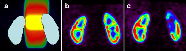
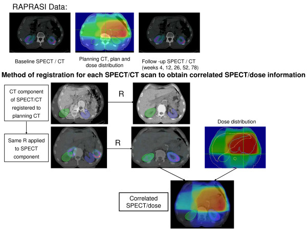
METHODS/DESIGN: The Radiotherapy of Abdomen with Precise Renal Assessment with SPECT/CT Imaging (RAPRASI) is an observational clinical research study with participating sites at Sir Charles Gairdner Hospital (SCGH) in Perth, Australia and the Peter MacCallum Cancer Centre (PMCC) in Melbourne, Australia. Eligible patients are those with upper gastrointestinal cancer, without metastatic disease, undergoing conformal radiotherapy that will involve incidental radiation to one or both kidneys. For each patient, total kidney function is being assessed before commencement of radiotherapy treatment and then at 4, 12, 26, 52 and 78 weeks after the first radiotherapy fraction, using two procedures: a Glomerular Filtration Rate (GFR) measurement using the 51Cr-ethylenediamine tetra-acetic acid (EDTA) clearance; and a regional kidney perfusion measurement assessing renal uptake of 99mTc-dimercaptosuccinic acid (DMSA), imaged with a Single Photon Emission Computed Tomography / Computed Tomography (SPECT/CT) system. The CT component of the SPECT/CT provides the anatomical reference of the kidney's position. The data is intended to reveal changes in regional kidney function over the study period after the radiotherapy. These SPECT/CT scans, co-registered with the radiotherapy treatment plan, will provide spatial correlation between the radiation dose and regional renal function as assessed by SPECT/CT. From this correlation, renal response patterns will likely be identified with the purpose of developing a predictive model.

TRIAL REGISTRATION

Australian New Zealand Clinical Trials Registry: ACTRN12609000322235.

摘要

背景

肾脏是上腹部癌症放射治疗的主要剂量限制器官。目前对肾脏辐射剂量反应的了解还很初步。更精确的剂量-体积反应模型,可使辐射剂量与肾脏功能的时空变化直接相关,这可能会改善上腹部肿瘤的放射治疗计划。

方法/设计:腹部放射治疗中精确肾评估的 SPECT/CT 成像(RAPRASI)是一项观察性临床研究,参与地点在澳大利亚珀斯的查尔斯·盖尔德纳爵士医院(SCGH)和澳大利亚墨尔本的彼得·麦卡伦癌症中心(PMCC)。符合条件的患者是患有上胃肠道癌、无转移疾病、接受适形放射治疗的患者,放射治疗会意外照射到一个或两个肾脏。对于每位患者,在开始放射治疗前以及在第一次放射治疗后 4、12、26、52 和 78 周,使用两种程序评估总肾功能:使用 51Cr-乙二胺四乙酸(EDTA)清除率测量肾小球滤过率(GFR);以及评估肾脏摄取 99mTc-二巯丁二酸(DMSA)的区域肾脏灌注测量,使用单光子发射计算机断层扫描/计算机断层扫描(SPECT/CT)系统进行成像。SPECT/CT 的 CT 组件提供了肾脏位置的解剖参考。该数据旨在揭示放射治疗后研究期间区域肾功能的变化。这些 SPECT/CT 扫描与放射治疗计划共同注册,将在 SPECT/CT 评估的区域肾功能与辐射剂量之间提供空间相关性。从这种相关性中,可能会确定肾脏反应模式,目的是开发预测模型。

试验注册

澳大利亚和新西兰临床试验注册中心:ACTRN12609000322235。

https://cdn.ncbi.nlm.nih.gov/pmc/blobs/4d7c/3751853/deedbb707279/1471-2407-13-381-2.jpg
https://cdn.ncbi.nlm.nih.gov/pmc/blobs/4d7c/3751853/dc7e6ae452c1/1471-2407-13-381-1.jpg
https://cdn.ncbi.nlm.nih.gov/pmc/blobs/4d7c/3751853/deedbb707279/1471-2407-13-381-2.jpg
https://cdn.ncbi.nlm.nih.gov/pmc/blobs/4d7c/3751853/dc7e6ae452c1/1471-2407-13-381-1.jpg
https://cdn.ncbi.nlm.nih.gov/pmc/blobs/4d7c/3751853/deedbb707279/1471-2407-13-381-2.jpg

相似文献

1
Radiotherapy of abdomen with precise renal assessment with SPECT/CT imaging (RAPRASI): design and methodology of a prospective trial to improve the understanding of kidney radiation dose response.腹部放疗中 SPECT/CT 成像的精确肾脏评估(RAPRASI):一项前瞻性试验的设计和方法,旨在提高对肾脏放射剂量反应的理解。
BMC Cancer. 2013 Aug 10;13:381. doi: 10.1186/1471-2407-13-381.
2
Dose-Effect Relationship of Kidney Function After SABR for Primary Renal Cell Carcinoma: TROG 15.03 FASTRACK II.SABR 治疗原发性肾细胞癌后肾功能的剂量-效应关系:TROG 15.03 FASTRACK II。
Int J Radiat Oncol Biol Phys. 2024 Nov 1;120(3):648-654. doi: 10.1016/j.ijrobp.2024.04.066. Epub 2024 Apr 26.
3
Short communication: timeline of radiation-induced kidney function loss after stereotactic ablative body radiotherapy of renal cell carcinoma as evaluated by serial (99m)Tc-DMSA SPECT/CT.简短通讯:通过连续(99m)Tc-DMSA SPECT/CT评估肾细胞癌立体定向消融放疗后放射性肾功能丧失的时间线
Radiat Oncol. 2014 Nov 26;9:253. doi: 10.1186/s13014-014-0253-z.
4
Determination of single-kidney glomerular filtration rate (GFR) with CT urography versus renal dynamic imaging Gates method.应用 CT 尿路造影与肾动态成像门控法测定单肾肾小球滤过率(GFR)。
Eur Radiol. 2018 Mar;28(3):1077-1084. doi: 10.1007/s00330-017-5061-z. Epub 2017 Oct 2.
5
Impact of stereotactic radiotherapy on kidney function in primary renal cell carcinoma: Establishing a dose-response relationship.立体定向放疗对原发性肾细胞癌肾功能的影响:建立剂量反应关系。
Radiother Oncol. 2016 Mar;118(3):540-6. doi: 10.1016/j.radonc.2016.01.027. Epub 2016 Feb 9.
6
Decreased Lung Perfusion After Breast/Chest Wall Irradiation: Quantitative Results From a Prospective Clinical Trial.乳房/胸壁放疗后肺灌注减少:一项前瞻性临床试验的定量结果
Int J Radiat Oncol Biol Phys. 2017 Feb 1;97(2):296-302. doi: 10.1016/j.ijrobp.2016.10.012. Epub 2016 Oct 19.
7
Renal SPECT/CT with 99mTc-dimercaptosuccinic acid is a non-invasive predictive marker for the development of interstitial fibrosis in a rat model of renal insufficiency.肾 SPECT/CT 联合 99mTc-二巯丁二酸检测对肾功能不全大鼠模型间质纤维化发展的预测作用
Nephrol Dial Transplant. 2021 Apr 26;36(5):804-810. doi: 10.1093/ndt/gfaa374.
8
(99m)Tc-DTPA dynamic SPECT and CT volumetry for measuring split renal function in live kidney donors.(99m)Tc-DTPA 动态 SPECT 和 CT 容积测量法在活体供肾者分肾功能测量中的应用。
Ann Nucl Med. 2010 Apr;24(3):189-95. doi: 10.1007/s12149-010-0349-y. Epub 2010 Mar 9.
9
Clinical utility of co-registered respiratory-gated( 99m)Tc-Technegas/MAA SPECT-CT images in the assessment of regional lung functional impairment in patients with lung cancer.联合配准呼吸门控(99m)Tc-锝气体/大颗粒聚合人血清白蛋白单光子发射计算机断层扫描/计算机断层扫描成像在评估肺癌患者局部肺功能损害中的临床应用
Eur J Nucl Med Mol Imaging. 2004 Sep;31(9):1280-90. doi: 10.1007/s00259-004-1558-1. Epub 2004 Jun 10.
10
Intensity modulated radiotherapy of non-small-cell lung cancer incorporating SPECT ventilation imaging.非小细胞肺癌的调强放疗结合 SPECT 通气显像。
Med Phys. 2010 Apr;37(4):1863-72. doi: 10.1118/1.3358128.

引用本文的文献

1
Novel radiopharmaceuticals for molecular imaging of renal cell carcinoma.用于肾细胞癌分子成像的新型放射性药物。
BMJ Oncol. 2025 Aug 17;4(1):e000645. doi: 10.1136/bmjonc-2024-000645. eCollection 2025.
2
Heterogeneity of absorbed dose distribution in kidney tissues and dose-response modelling of nephrotoxicity in radiopharmaceutical therapy with beta-particle emitters: A review.β粒子发射放射性药物治疗中肾组织内吸收剂量分布的异质性与肾毒性的剂量反应建模:综述。
Z Med Phys. 2024 Nov;34(4):491-509. doi: 10.1016/j.zemedi.2023.02.006. Epub 2023 Apr 6.
3
A novel rotation method with variable-angle anterior probe for correcting the depth of the kidney to optimize renal dynamic imaging.

本文引用的文献

1
Imaging radiation-induced normal tissue injury.影像学诊断辐射导致的正常组织损伤。
Radiat Res. 2012 Apr;177(4):449-66. doi: 10.1667/rr2530.1. Epub 2012 Feb 21.
2
Mechanistic simulation of normal-tissue damage in radiotherapy--implications for dose-volume analyses.放射治疗中正常组织损伤的机制模拟——对剂量-体积分析的影响。
Phys Med Biol. 2010 Apr 21;55(8):2121-36. doi: 10.1088/0031-9155/55/8/001. Epub 2010 Mar 19.
3
Radiation-associated kidney injury.辐射相关性肾损伤。
一种采用可变角度前探头的新型旋转方法,用于校正肾脏深度以优化肾动态成像。
EJNMMI Phys. 2022 Nov 17;9(1):79. doi: 10.1186/s40658-022-00511-w.
4
Regional Responses in Radiation-Induced Normal Tissue Damage.辐射诱导的正常组织损伤中的区域反应
Cancers (Basel). 2021 Jan 20;13(3):367. doi: 10.3390/cancers13030367.
5
Inhibition of mTORC1 signaling protects kidney from irradiation-induced toxicity via accelerating recovery of renal stem-like cells.mTORC1 信号抑制通过加速肾脏干细胞样细胞的恢复来保护肾脏免受辐射诱导的毒性。
Stem Cell Res Ther. 2018 Aug 14;9(1):219. doi: 10.1186/s13287-018-0963-5.
Int J Radiat Oncol Biol Phys. 2010 Mar 1;76(3 Suppl):S108-15. doi: 10.1016/j.ijrobp.2009.02.089.
4
Hybrid imaging (SPECT/CT and PET/CT)--improving the diagnostic accuracy of functional/metabolic and anatomic imaging.混合成像(SPECT/CT 和 PET/CT)——提高功能/代谢和解剖成像的诊断准确性。
Semin Nucl Med. 2009 Jul;39(4):264-75. doi: 10.1053/j.semnuclmed.2009.03.004.
5
Use of the BrainLAB ExacTrac X-Ray 6D system in image-guided radiotherapy.BrainLAB ExacTrac X射线6D系统在图像引导放射治疗中的应用。
Med Dosim. 2008 Summer;33(2):124-34. doi: 10.1016/j.meddos.2008.02.005. Epub 2008 Apr 1.
6
Using an onboard kilovoltage imager to measure setup deviation in intensity-modulated radiation therapy for head-and-neck patients.使用机载千伏成像仪测量头颈部患者调强放射治疗中的摆位偏差。
J Appl Clin Med Phys. 2007 Sep 24;8(4):28-44. doi: 10.1120/jacmp.v8i4.2439.
7
3D radiotherapy can be safely combined with sandwich systemic gemcitabine chemotherapy in the management of pancreatic cancer: factors influencing outcome.在胰腺癌的治疗中,三维放射治疗可安全地与夹心式吉西他滨全身化疗联合使用:影响疗效的因素
Int J Radiat Oncol Biol Phys. 2008 Apr 1;70(5):1438-46. doi: 10.1016/j.ijrobp.2007.08.070. Epub 2007 Dec 31.
8
A four-dimensional CT-based evaluation of techniques for gastric irradiation.基于四维CT的胃部放疗技术评估
Int J Radiat Oncol Biol Phys. 2007 Nov 1;69(3):903-9. doi: 10.1016/j.ijrobp.2007.06.062.
9
Interfractional variations in patient setup and anatomic change assessed by daily computed tomography.通过每日计算机断层扫描评估患者摆位和解剖结构变化的分次间差异。
Int J Radiat Oncol Biol Phys. 2007 Jun 1;68(2):581-91. doi: 10.1016/j.ijrobp.2006.12.024. Epub 2007 Feb 27.
10
Prospective study on late renal toxicity following postoperative chemoradiotherapy in gastric cancer.胃癌术后放化疗后迟发性肾毒性的前瞻性研究
Int J Radiat Oncol Biol Phys. 2007 Mar 1;67(3):781-5. doi: 10.1016/j.ijrobp.2006.09.012. Epub 2006 Dec 8.