Center for Medical Image Science and Visualization, CMIV, Linköping University, Linköping, Sweden.
PLoS One. 2013 Aug 5;8(8):e70864. doi: 10.1371/journal.pone.0070864. Print 2013.
To present a method for generating reference maps of typical brain characteristics of groups of subjects using a novel combination of rapid quantitative Magnetic Resonance Imaging (qMRI) and brain normalization. The reference maps can be used to detect significant tissue differences in patients, both locally and globally.
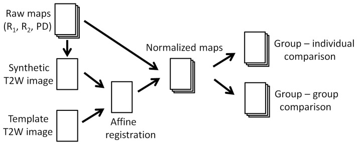
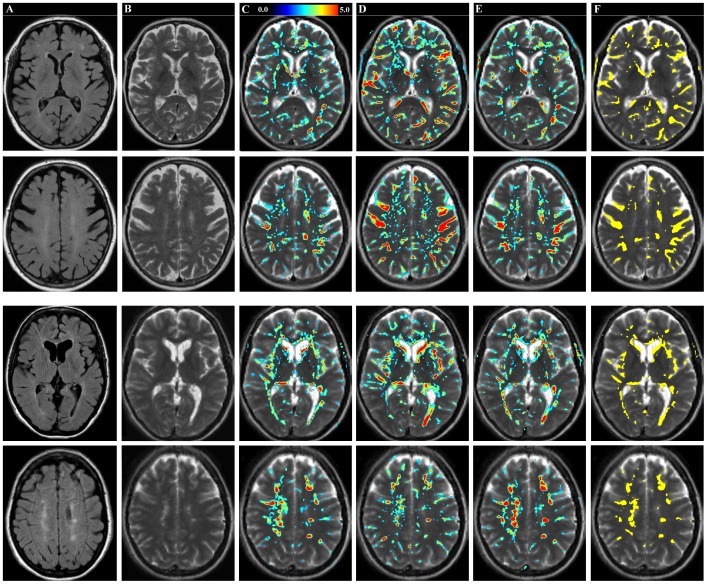
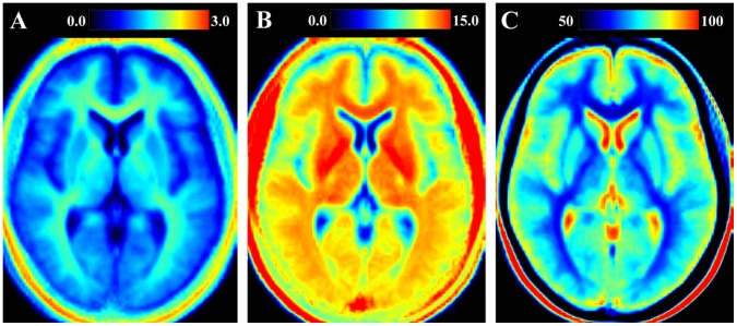
A rapid qMRI method was used to obtain the longitudinal relaxation rate (R1), the transverse relaxation rate (R2) and the proton density (PD). These three tissue properties were measured in the brains of 32 healthy subjects and in one patient diagnosed with Multiple Sclerosis (MS). The maps were normalized to a standard brain template using a linear affine registration. The differences of the mean value ofR1, R2 and PD of 31 healthy subjects in comparison to the oldest healthy subject and in comparison to an MS patient were calculated. Larger anatomical structures were characterized using a standard atlas. The vector sum of the normalized differences was used to show significant tissue differences.
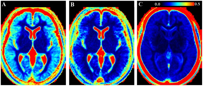
The coefficient of variation of the reference maps was high at the edges of the brain and the ventricles, moderate in the cortical grey matter and low in white matter and the deep grey matter structures. The elderly subject mainly showed significantly lower R1 and R2 and higher PD values along all sulci. The MS patient showed significantly lower R1 and R2 and higher PD values at the edges of the ventricular system as well as throughout the periventricular white matter, at the internal and external capsules and at each of the MS lesions.
Brain normalization of rapid qMRI is a promising new method to generate reference maps of typical brain characteristics and to automatically detect deviating tissue properties in the brain.
提出一种使用新型快速定量磁共振成像(qMRI)和脑归一化的组合生成典型脑特征的参考图谱的方法。参考图谱可用于局部和全局检测患者的显著组织差异。
使用快速 qMRI 方法获取 32 名健康受试者和 1 名多发性硬化症(MS)患者的脑内纵向弛豫率(R1)、横向弛豫率(R2)和质子密度(PD)。使用线性仿射配准将图谱归一化为标准脑模板。计算 31 名健康受试者相对于最年长的健康受试者和相对于 MS 患者的 R1、R2 和 PD 的平均值差异。使用标准图谱对较大的解剖结构进行特征描述。归一化差异的向量和用于显示显著的组织差异。
参考图谱的变异系数在脑和脑室边缘较高,在皮质灰质中中等,在白质和深部灰质结构中较低。年长的受试者主要沿所有脑沟显示出显著较低的 R1 和 R2 值和较高的 PD 值。MS 患者在脑室系统边缘以及整个脑室周围白质、内囊和外囊以及每个 MS 病变处均显示出显著较低的 R1 和 R2 值和较高的 PD 值。
快速 qMRI 的脑归一化是生成典型脑特征参考图谱并自动检测脑内偏离组织特性的一种很有前途的新方法。