NeuroCure Clinical Research Center and Experimental and Clinical Research Center, Charité-Universitätsmedizin Berlin and Max Delbrück Center for Molecular Medicine, Berlin, Germany.
PLoS One. 2013 Aug 6;8(8):e71145. doi: 10.1371/journal.pone.0071145. Print 2013.
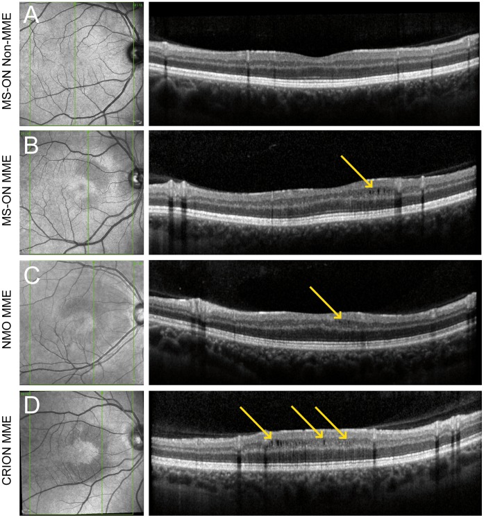
Microcystic macular edema (MME) and inner nuclear layer thickening (INL) were described in multiple sclerosis (MS) and neuromyelitis optica (NMO) patients using optical coherence tomography (OCT). The cause of these findings is currently unknown and a relation to inflammatory or degenerative processes in the optic nerve is discussed.
The aim of our study was to investigate whether INL thickening and MME are related to optic neuritis (ON) in various neuro-inflammatory disorders causingON: MS, NMO and chronic inflammatory optic neuropathy.
We retrospectively analyzed data from 216 MS patients, 39 patients with a clinically isolated syndrome, 20 NMO spectrum disorder patients, 9 patients with chronic inflammatory optic neuropathy and 121 healthy subjects. Intra-retinal layer segmentation was performed for the eyes of patients with unilateral ON. Scanning laser ophthalmoscopy (SLO) images were reviewed for characteristic ocular fundus changes.
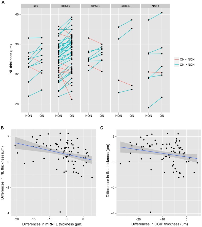
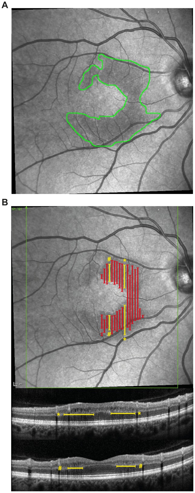
Intra-retinal layer segmentation showed that eyes with a history of ON displayed MME independent INL thickening compared to contralateral eyes without previous ON. MME was detected in 22 eyes from 15 patients (5.3% of all screened patients), including 7 patients with bilateral edema. Of these, 21 had a prior history of ON (95%). The SLO images of all 22 MME-affected eyes showed crescent-shaped texture changes which were visible in the perifoveal region. A second grader who was blinded to the results of the OCT classified all SLO images for the presence of these characteristic fundus changes. All MME eyes were correctly classified (sensitivity = 100%) with high specificity (95.2%).
This study shows that both MME and INL thickening occur in various neuro-inflammatory disorders associated with ON. We also demonstrate that detection and analysis of MME by OCT is not limited to B-scans, but also possible using SLO images.
多发性硬化症(MS)和视神经脊髓炎(NMO)患者的光学相干断层扫描(OCT)显示出微囊样黄斑水肿(MME)和内核层增厚(INL)。目前尚不清楚这些发现的原因,有人认为它们与视神经炎症或退行性过程有关。
本研究旨在探讨各种引起视神经炎(ON)的神经炎症性疾病中,INL 增厚和 MME 是否与 MS、NMO 和慢性炎症性视神经病变中的 ON 有关。
我们回顾性分析了 216 例 MS 患者、39 例临床孤立综合征患者、20 例 NMO 谱系障碍患者、9 例慢性炎症性视神经病变患者和 121 例健康对照者的数据。对单侧 ON 患者的眼部进行视网膜内部分层分析。对眼底检查的特征性改变进行扫激光检眼镜(SLO)图像复查。
视网膜内部分层分析显示,有 ON 病史的眼与对侧无 ON 病史的眼相比,存在 MME 伴发的 INL 增厚。在 15 例患者的 22 只眼中检测到 MME(所有筛查患者的 5.3%),其中 7 例为双侧水肿。这些患者中,21 例有 ON 病史(95%)。所有 22 只 MME 受累眼的 SLO 图像均显示出新月形纹理改变,这些改变在中心凹旁可见。一位对 OCT 结果不知情的二级评估员对这些特征性眼底改变的存在情况对所有 SLO 图像进行了分类。所有 MME 眼均被正确分类(敏感性为 100%),特异性高(95.2%)。
本研究表明,MME 和 INL 增厚均发生在与 ON 相关的各种神经炎症性疾病中。我们还证明,通过 OCT 检测和分析 MME 不仅限于 B 扫描,还可以使用 SLO 图像进行。