Graduate Program of Biomedical Engineering, Duke University, Durham, NC, USA.
Int J Hyperthermia. 2013 Sep;29(6):569-81. doi: 10.3109/02656736.2013.790091.
During hyperthermia (HT), the therapeutic response of tumours varies substantially within the target temperature range (39-43 °C). Current thermometry methods are either invasive or measure only temperature change, which limits the ability to study tissue responses to HT. This study combines manganese-containing low temperature sensitive liposomes (Mn-LTSL) with proton resonance frequency shift (PRFS) thermometry to measure absolute temperature in tumours with high spatial and temporal resolution using MRI.
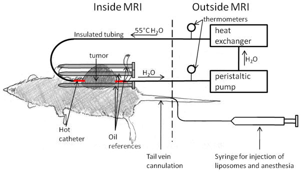
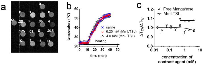
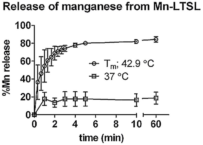
Liposomes were loaded with 300 mM MnSO(4). The phase transition temperature (T(m)) of Mn-LTSL samples was measured by differential scanning calorimetry (DSC). The release of manganese from Mn-LTSL in saline was characterised with inductively coupled plasma atomic emission spectroscopy. A 2T GE small animal scanner was used to acquire dynamic T1-weighted images and temperature change images of Mn-LTSL in saline phantoms and fibrosarcoma-bearing Fisher-344 rats receiving hyperthermia after Mn-LTSL injection.
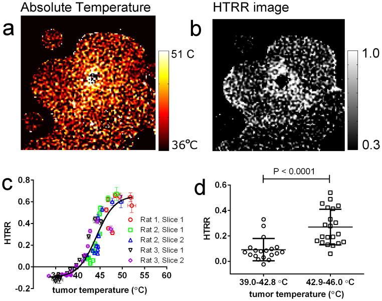
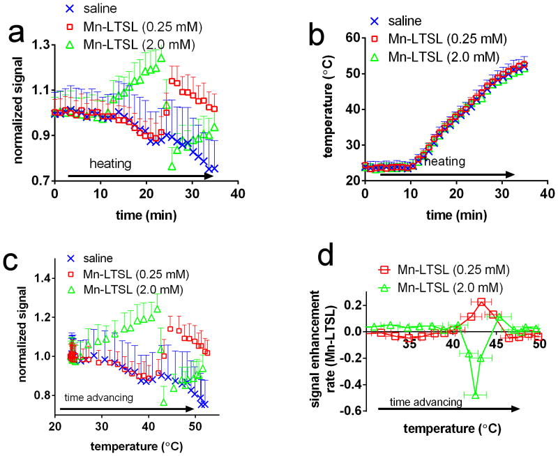
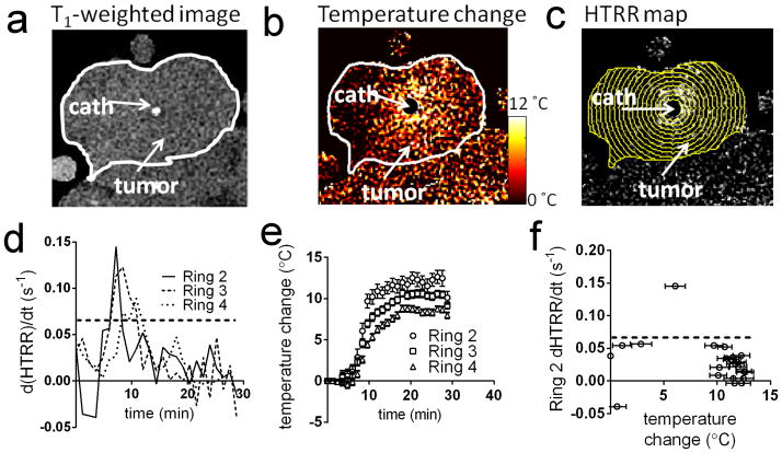
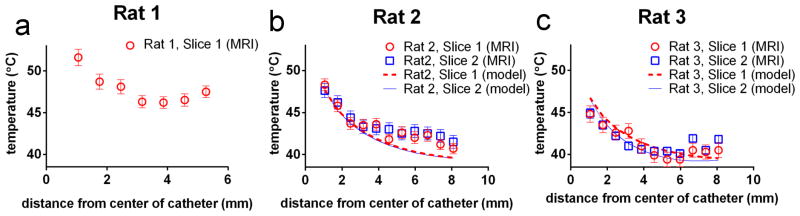
The T(m) of Mn-LTSL in rat blood was 42.9 ± 0.2 °C (DSC). For Mn-LTSL samples (0.06 mM-0.5 mM Mn(2+) in saline) heated monotonically from 30 °C to 50 °C, a peak in the rate of MRI signal enhancement occurred at 43.1° ± 0.3 °C. The same peak in signal enhancement rate was observed during heating of fibrosarcoma tumours (N = 3) after injection of Mn-LTSL, and the peak was used to convert temperature change images into absolute temperature. Accuracies of calibrated temperature measurements were in the range 0.9-1.8 °C.
The release of Mn(2+) from Mn-LTSL affects the rate of MR signal enhancement which enables conversion of MRI-based temperature change images to absolute temperature.
在高热(HT)期间,肿瘤的治疗反应在目标温度范围内(39-43°C)有很大差异。当前的测温方法要么具有侵入性,要么只能测量温度变化,这限制了研究组织对 HT 的反应的能力。本研究结合含锰低温敏感脂质体(Mn-LTSL)和质子共振频率偏移(PRFS)测温法,使用 MRI 以高空间和时间分辨率测量肿瘤中的绝对温度。
脂质体负载 300mM MnSO4。Mn-LTSL 样品的相变温度(T(m))通过差示扫描量热法(DSC)测量。Mn-LTSL 在盐水中释放锰的情况通过电感耦合等离子体原子发射光谱法进行了表征。使用 2T GE 小动物扫描仪在盐水体模和接受 Mn-LTSL 注射后接受 HT 的纤维肉瘤荷瘤 Fisher-344 大鼠中获取 Mn-LTSL 的动态 T1 加权图像和温度变化图像。
Mn-LTSL 在大鼠血液中的 T(m)为 42.9±0.2°C(DSC)。对于从 30°C 单调加热至 50°C 的 Mn-LTSL 样品(0.06mM-0.5mM Mn(2+)在盐水中),在 43.1°±0.3°C 处出现 MRI 信号增强率的峰值。在注射 Mn-LTSL 后,纤维肉瘤肿瘤(N=3)的加热过程中也观察到相同的信号增强率峰值,该峰值用于将温度变化图像转换为绝对温度。校准温度测量的精度在 0.9-1.8°C 范围内。
Mn-LTSL 中 Mn(2+)的释放会影响 MRI 信号增强率,从而可以将基于 MRI 的温度变化图像转换为绝对温度。