• 文献检索
  • 文档翻译
  • 深度研究
  • 学术资讯
  • Suppr Zotero 插件Zotero 插件
  • 邀请有礼
  • 套餐&价格
  • 历史记录
应用&插件
Suppr Zotero 插件Zotero 插件浏览器插件Mac 客户端Windows 客户端微信小程序
定价
高级版会员购买积分包购买API积分包
服务
文献检索文档翻译深度研究API 文档MCP 服务
关于我们
关于 Suppr公司介绍联系我们用户协议隐私条款
关注我们

Suppr 超能文献

核心技术专利:CN118964589B侵权必究
粤ICP备2023148730 号-1Suppr @ 2026

文献检索

告别复杂PubMed语法,用中文像聊天一样搜索,搜遍4000万医学文献。AI智能推荐,让科研检索更轻松。

立即免费搜索

文件翻译

保留排版,准确专业,支持PDF/Word/PPT等文件格式,支持 12+语言互译。

免费翻译文档

深度研究

AI帮你快速写综述,25分钟生成高质量综述,智能提取关键信息,辅助科研写作。

立即免费体验

心脏 CT 对冠状动脉疾病的生理学评估。

Physiologic assessment of coronary artery disease by cardiac computed tomography.

机构信息

Division of Cardiology, Kaiser Permanente, Panorama City, CA, USA.

出版信息

Korean Circ J. 2013 Jul;43(7):435-42. doi: 10.4070/kcj.2013.43.7.435.

DOI:10.4070/kcj.2013.43.7.435
PMID:23964289
原文链接:https://pmc.ncbi.nlm.nih.gov/articles/PMC3744730/
Abstract

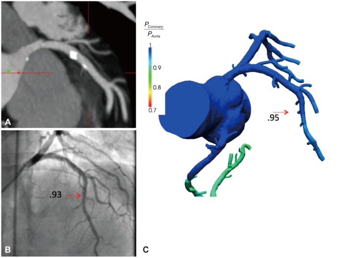
Coronary artery disease (CAD) remains the leading cause of death and morbidity worldwide. To date, diagnostic evaluation of patients with suspected CAD has relied upon the use of physiologic non-invasive testing by stress electrocardiography, echocardiography, myocardial perfusion imaging (MPI) and magnetic resonance imaging. Indeed, the importance of physiologic evaluation of CAD has been highlighted by large-scale randomized trials that demonstrate the propitious benefit of an integrated anatomic-physiologic evaluation method by performing lesion-specific ischemia assessment by fractional flow reserve (FFR)-widely considered the "gold" standard for ischemia assessment-at the time of invasive angiography. Coronary CT angiography (CCTA) has emerged as an attractive non-invasive test for anatomic illustration of the coronary arteries and atherosclerotic plaque. In a series of prospective multicenter trials, CCTA has been proven as having high diagnostic performance for stenosis detection as compared to invasive angiography. Nevertheless, CCTA evaluation of obstructive stenoses is prone to overestimation of severity and further, detection of stenoses by CCTA does not reliably determine the hemodynamic significance of the visualized lesions. Recently, a series of technological innovations have advanced the possibility of CCTA to enable physiologic evaluation of CAD, thereby creating the potential of this test to provide an integrated anatomic-physiologic assessment of CAD. These advances include rest-stress MPI by CCTA as well as the use of computational fluid dynamics to non-invasively calculate FFR from a typically acquired CCTA. The purpose of this review is to summarize the most recent data addressing these 2 physiologic methods of CAD evaluation by CCTA.

摘要

冠心病(CAD)仍然是全球范围内导致死亡和发病的主要原因。迄今为止,疑似 CAD 患者的诊断评估依赖于通过应激心电图、超声心动图、心肌灌注成像(MPI)和磁共振成像进行的生理非侵入性测试。实际上,大规模随机试验强调了生理评估 CAD 的重要性,这些试验表明通过在血管造影时进行特定病变的缺血评估(广泛认为是缺血评估的“金标准”)来进行病变特异性缺血评估的综合解剖生理评估方法具有有利的益处。冠状动脉 CT 血管造影(CCTA)已成为一种有吸引力的非侵入性测试,用于冠状动脉和动脉粥样硬化斑块的解剖描述。在一系列前瞻性多中心试验中,CCTA 已被证明在狭窄检测方面具有比血管造影更高的诊断性能。然而,CCTA 对阻塞性狭窄的评估容易高估狭窄程度,而且,CCTA 检测到的狭窄并不能可靠地确定可视化病变的血流动力学意义。最近,一系列技术创新提高了 CCTA 进行 CAD 生理评估的可能性,从而使该测试有可能提供 CAD 的综合解剖生理评估。这些进展包括 CCTA 进行静息-应激 MPI 以及使用计算流体动力学从典型获得的 CCTA 无创计算 FFR。本综述的目的是总结最新的数据,这些数据涉及 CCTA 评估 CAD 的这两种生理方法。

https://cdn.ncbi.nlm.nih.gov/pmc/blobs/115f/3744730/20872497de19/kcj-43-435-g002.jpg
https://cdn.ncbi.nlm.nih.gov/pmc/blobs/115f/3744730/646e91334d00/kcj-43-435-g001.jpg
https://cdn.ncbi.nlm.nih.gov/pmc/blobs/115f/3744730/20872497de19/kcj-43-435-g002.jpg
https://cdn.ncbi.nlm.nih.gov/pmc/blobs/115f/3744730/646e91334d00/kcj-43-435-g001.jpg
https://cdn.ncbi.nlm.nih.gov/pmc/blobs/115f/3744730/20872497de19/kcj-43-435-g002.jpg

相似文献

1
Physiologic assessment of coronary artery disease by cardiac computed tomography.心脏 CT 对冠状动脉疾病的生理学评估。
Korean Circ J. 2013 Jul;43(7):435-42. doi: 10.4070/kcj.2013.43.7.435.
2
Noninvasive diagnosis of ischemia-causing coronary stenosis using CT angiography: diagnostic value of transluminal attenuation gradient and fractional flow reserve computed from coronary CT angiography compared to invasively measured fractional flow reserve.采用 CT 血管造影术无创诊断缺血性冠状动脉狭窄:血管内测量的血流储备分数与 CT 血管造影术计算的管腔衰减梯度和血流储备分数的诊断价值比较。
JACC Cardiovasc Imaging. 2012 Nov;5(11):1088-96. doi: 10.1016/j.jcmg.2012.09.002.
3
Diagnosis of ischemia-causing coronary stenoses by noninvasive fractional flow reserve computed from coronary computed tomographic angiograms. Results from the prospective multicenter DISCOVER-FLOW (Diagnosis of Ischemia-Causing Stenoses Obtained Via Noninvasive Fractional Flow Reserve) study.通过冠状动脉计算机断层血管造影计算无创性血流储备分数诊断缺血性冠状动脉狭窄。前瞻性多中心 DISCOVER-FLOW(通过无创性血流储备分数诊断缺血性狭窄)研究结果。
J Am Coll Cardiol. 2011 Nov 1;58(19):1989-97. doi: 10.1016/j.jacc.2011.06.066.
4
Prospective comparison of integrated on-site CT-fractional flow reserve and static CT perfusion with coronary CT angiography for detection of flow-limiting coronary stenosis.前瞻性比较整合的 CT 现场分段血流储备分数和 CT 灌注与冠状动脉 CT 血管造影用于检测限制血流的冠状动脉狭窄。
Eur Radiol. 2021 Jul;31(7):5096-5105. doi: 10.1007/s00330-020-07508-y. Epub 2021 Jan 6.
5
Rationale and Design of the CREDENCE Trial: computed TomogRaphic evaluation of atherosclerotic DEtermiNants of myocardial IsChEmia.CREDENCE试验的原理与设计:心肌缺血的动脉粥样硬化决定因素的计算机断层扫描评估
BMC Cardiovasc Disord. 2016 Oct 6;16(1):190. doi: 10.1186/s12872-016-0360-x.
6
Diagnostic performance of transluminal attenuation gradient and fractional flow reserve by coronary computed tomographic angiography (FFR(CT)) compared to invasive FFR: a sub-group analysis from the DISCOVER-FLOW and DeFACTO studies.与有创血流储备分数(FFR)相比,冠状动脉计算机断层扫描血管造影术(FFR(CT))的腔内衰减梯度和血流储备分数的诊断性能:来自DISCOVER-FLOW和DeFACTO研究的亚组分析。
Int J Cardiovasc Imaging. 2015 Aug;31(6):1251-9. doi: 10.1007/s10554-015-0666-2. Epub 2015 Apr 24.
7
Rationale and design of advantage (additional diagnostic value of CT perfusion over coronary CT angiography in stented patients with suspected in-stent restenosis or coronary artery disease progression) prospective study.优势(CT 灌注相对于 CT 血管造影在支架置入患者中诊断支架内再狭窄或冠状动脉疾病进展的附加价值)前瞻性研究的原理和设计。
J Cardiovasc Comput Tomogr. 2018 Sep-Oct;12(5):411-417. doi: 10.1016/j.jcct.2018.06.003. Epub 2018 Jun 18.
8
[Non-invasive functional assessment of coronary stenosis by cardiac computed tomography].[心脏计算机断层扫描对冠状动脉狭窄的无创功能评估]
G Ital Cardiol (Rome). 2019 Jul-Aug;20(7):417-428. doi: 10.1714/3190.31685.
9
The New Frontier of Cardiac Computed Tomography Angiography: Fractional Flow Reserve and Stress Myocardial Perfusion.心脏计算机断层扫描血管造影的新前沿:血流储备分数和负荷心肌灌注
Curr Treat Options Cardiovasc Med. 2016 Dec;18(12):74. doi: 10.1007/s11936-016-0493-3.
10
A study of noninvasive fractional flow reserve derived from a simplified method based on coronary computed tomography angiography in suspected coronary artery disease.一项关于基于冠状动脉计算机断层扫描血管造影的简化方法得出的无创血流储备分数在疑似冠状动脉疾病中的研究。
Biomed Eng Online. 2017 Apr 14;16(1):43. doi: 10.1186/s12938-017-0330-2.

引用本文的文献

1
Color-coded duplex sonography vs. 3.0 Tesla magnetic resonance angiography for detection of intracranial stenosis of the internal carotid artery: A prospective cohort study.彩色编码双功超声与3.0特斯拉磁共振血管造影术用于检测颈内动脉颅内狭窄:一项前瞻性队列研究。
Exp Ther Med. 2020 Jan;19(1):473-480. doi: 10.3892/etm.2019.8255. Epub 2019 Nov 29.
2
Physiologic Assessment of Coronary Artery Disease: Focus on Fractional Flow Reserve.冠状动脉疾病的生理学评估:聚焦于血流储备分数
Korean J Radiol. 2016 May-Jun;17(3):307-20. doi: 10.3348/kjr.2016.17.3.307. Epub 2016 Apr 14.
3
Direct comparison of stress- and rest-dual-energy computed tomography for detection of myocardial perfusion defect.

本文引用的文献

1
Dual-energy CT and its potential use for quantitative myocardial CT perfusion.双能 CT 及其在定量心肌 CT 灌注中的潜在应用。
J Cardiovasc Comput Tomogr. 2012 Sep-Oct;6(5):308-17. doi: 10.1016/j.jcct.2012.07.002. Epub 2012 Aug 24.
2
Fractional flow reserve-guided PCI versus medical therapy in stable coronary disease.临界血流储备分数指导下的经皮冠状动脉介入治疗与稳定型冠心病的药物治疗。
N Engl J Med. 2012 Sep 13;367(11):991-1001. doi: 10.1056/NEJMoa1205361. Epub 2012 Aug 27.
3
Diagnostic accuracy of fractional flow reserve from anatomic CT angiography.
直接比较应激和静息双能 CT 检测心肌灌注缺损。
Int J Cardiovasc Imaging. 2014 Jun;30 Suppl 1:41-53. doi: 10.1007/s10554-014-0410-3. Epub 2014 Apr 3.
解剖 CT 血管造影中血流储备分数的诊断准确性。
JAMA. 2012 Sep 26;308(12):1237-45. doi: 10.1001/2012.jama.11274.
4
Evaluation of myocardial CT perfusion in patients presenting with acute chest pain to the emergency department: comparison with SPECT-myocardial perfusion imaging.评价以急性胸痛为主诉就诊于急诊科患者的心肌 CT 灌注:与 SPECT 心肌灌注成像比较。
Heart. 2012 Oct;98(20):1510-7. doi: 10.1136/heartjnl-2012-302531. Epub 2012 Aug 15.
5
CT myocardial perfusion: a step towards quantification.CT心肌灌注:迈向量化的一步。
Heart. 2012 Apr;98(7):521-2. doi: 10.1136/heartjnl-2012-301677.
6
Quantitative myocardial CT perfusion: a pictorial review and the current state of technology development.定量心肌 CT 灌注:影像学综述与当前技术发展状况。
J Cardiovasc Comput Tomogr. 2011 Nov-Dec;5(6):467-81. doi: 10.1016/j.jcct.2011.11.002. Epub 2011 Nov 12.
7
Diagnosis of ischemia-causing coronary stenoses by noninvasive fractional flow reserve computed from coronary computed tomographic angiograms. Results from the prospective multicenter DISCOVER-FLOW (Diagnosis of Ischemia-Causing Stenoses Obtained Via Noninvasive Fractional Flow Reserve) study.通过冠状动脉计算机断层血管造影计算无创性血流储备分数诊断缺血性冠状动脉狭窄。前瞻性多中心 DISCOVER-FLOW(通过无创性血流储备分数诊断缺血性狭窄)研究结果。
J Am Coll Cardiol. 2011 Nov 1;58(19):1989-97. doi: 10.1016/j.jacc.2011.06.066.
8
Adenosine stress high-pitch 128-slice dual-source myocardial computed tomography perfusion for imaging of reversible myocardial ischemia: comparison with magnetic resonance imaging.腺苷负荷高心率 128 层双源 CT 心肌灌注成像诊断可逆性心肌缺血:与磁共振成像比较。
Circ Cardiovasc Imaging. 2011 Sep;4(5):540-9. doi: 10.1161/CIRCIMAGING.110.961250. Epub 2011 Aug 23.
9
Forecasting the future of cardiovascular disease in the United States: a policy statement from the American Heart Association.预测美国心血管疾病的未来:美国心脏协会的政策声明。
Circulation. 2011 Mar 1;123(8):933-44. doi: 10.1161/CIR.0b013e31820a55f5. Epub 2011 Jan 24.
10
Heart disease and stroke statistics--2011 update: a report from the American Heart Association.心脏病和中风统计数据--2011 年更新:来自美国心脏协会的报告。
Circulation. 2011 Feb 1;123(4):e18-e209. doi: 10.1161/CIR.0b013e3182009701. Epub 2010 Dec 15.