Institute of Genetic Medicine, Newcastle University, Newcastle upon Tyne, United Kingdom.
PLoS One. 2013 Aug 14;8(8):e70993. doi: 10.1371/journal.pone.0070993. eCollection 2013.
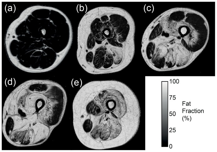
Outcome measures for clinical trials in neuromuscular diseases are typically based on physical assessments which are dependent on patient effort, combine the effort of different muscle groups, and may not be sensitive to progression over short trial periods in slow-progressing diseases. We hypothesised that quantitative fat imaging by MRI (Dixon technique) could provide more discriminating quantitative, patient-independent measurements of the progress of muscle fat replacement within individual muscle groups.
To determine whether quantitative fat imaging could measure disease progression in a cohort of limb-girdle muscular dystrophy 2I (LGMD2I) patients over a 12 month period.
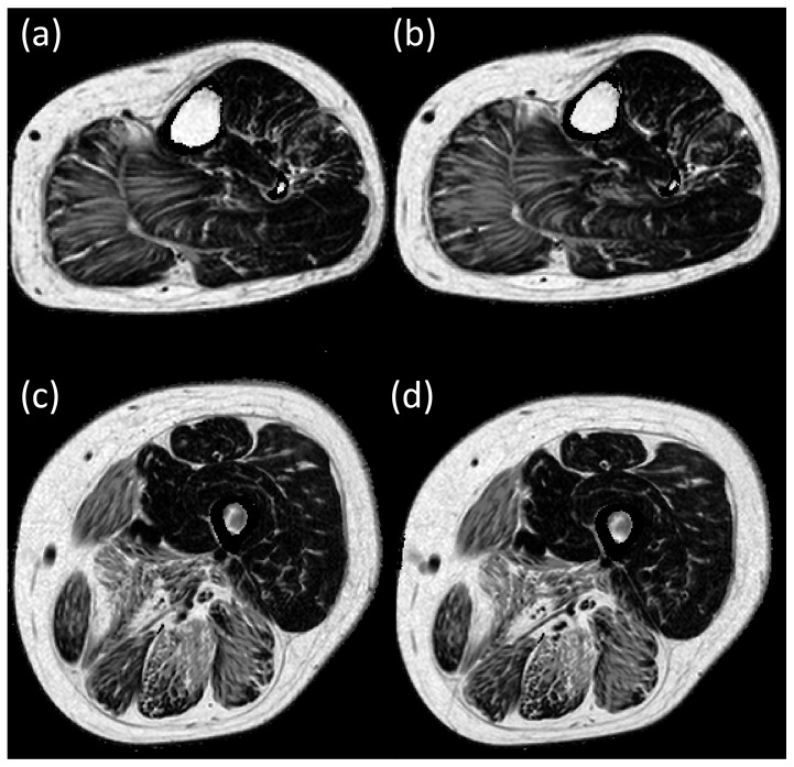
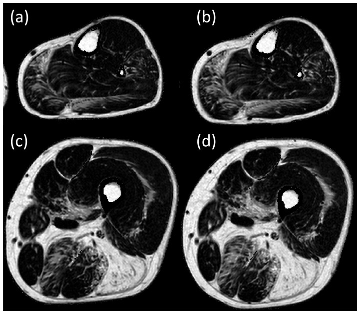
32 adult patients (17 male;15 female) from 4 European tertiary referral centres with the homozygous c.826C>A mutation in the fukutin-related protein gene (FKRP) completed baseline and follow up measurements 12 months later. Quantitative fat imaging was performed and muscle fat fraction change was compared with (i) muscle strength and function assessed using standardized physical tests and (ii) standard T1-weighted MRI graded on a 6 point scale.
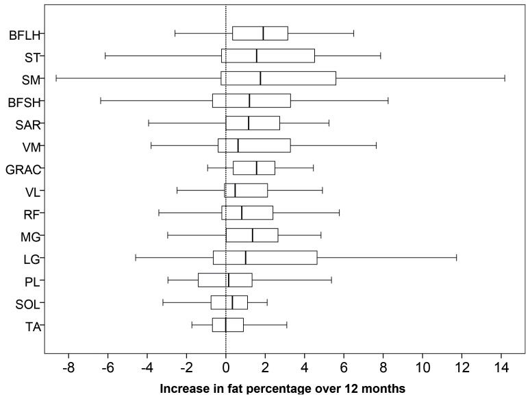
There was a significant increase in muscle fat fraction in 9 of the 14 muscles analyzed using the quantitative MRI technique from baseline to 12 months follow up. Changes were not seen in the conventional longitudinal physical assessments or in qualitative scoring of the T₁w images.
Quantitative muscle MRI, using the Dixon technique, could be used as an important longitudinal outcome measure to assess muscle pathology and monitor therapeutic efficacy in patients with LGMD2I.
神经肌肉疾病临床试验的结果测量通常基于物理评估,这些评估依赖于患者的努力,综合了不同肌肉群的努力,并且在进展缓慢的疾病的短期试验期间可能对进展不敏感。我们假设磁共振成像(Dixon 技术)的定量脂肪成像可以提供更具辨别力的、独立于患者的个体肌肉群中肌肉脂肪替代进展的定量测量。
确定定量脂肪成像是否可以在 12 个月的时间内测量肢带型肌肉营养不良 2I(LGMD2I)患者队列的疾病进展。
来自 4 个欧洲三级转诊中心的 32 名成年患者(17 名男性;15 名女性)携带 FKRP 基因的纯合 c.826C>A 突变,在基线和 12 个月后完成随访测量。进行定量脂肪成像,并比较肌肉脂肪分数变化与(i)使用标准化物理测试评估的肌肉力量和功能,以及(ii)标准 T1 加权 MRI 按 6 分制分级。
在使用定量 MRI 技术分析的 14 块肌肉中有 9 块肌肉的肌肉脂肪分数从基线到 12 个月的随访期间显著增加。在常规纵向物理评估或 T₁w 图像的定性评分中未观察到变化。
使用 Dixon 技术的定量肌肉 MRI 可作为评估肌肉病理和监测 LGMD2I 患者治疗效果的重要纵向结果测量方法。