MOE Key Laboratory of Aquatic Product Safety/State Key Laboratory of Biocontrol, School of Life Sciences, Sun Yat-Sen University, Guangzhou, People's Republic of China.
PLoS One. 2013 Aug 14;8(8):e72592. doi: 10.1371/journal.pone.0072592. eCollection 2013.
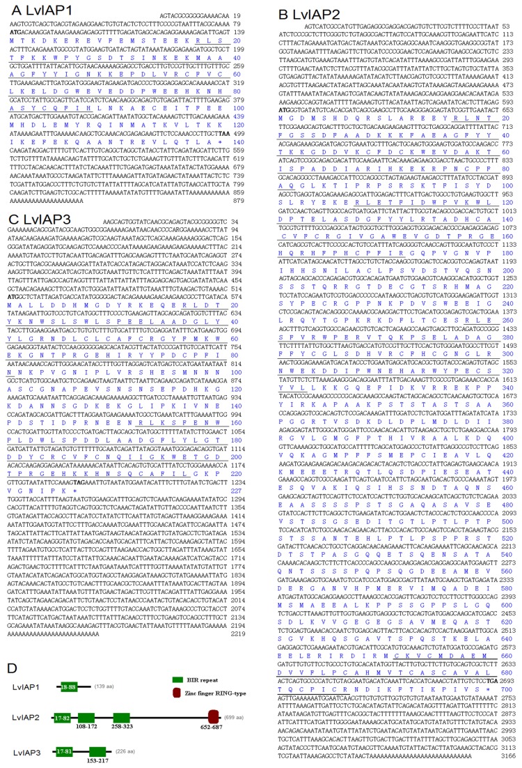
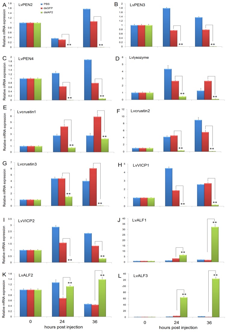
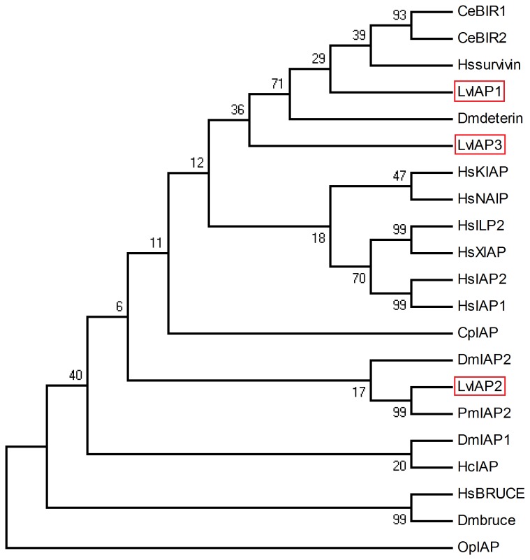
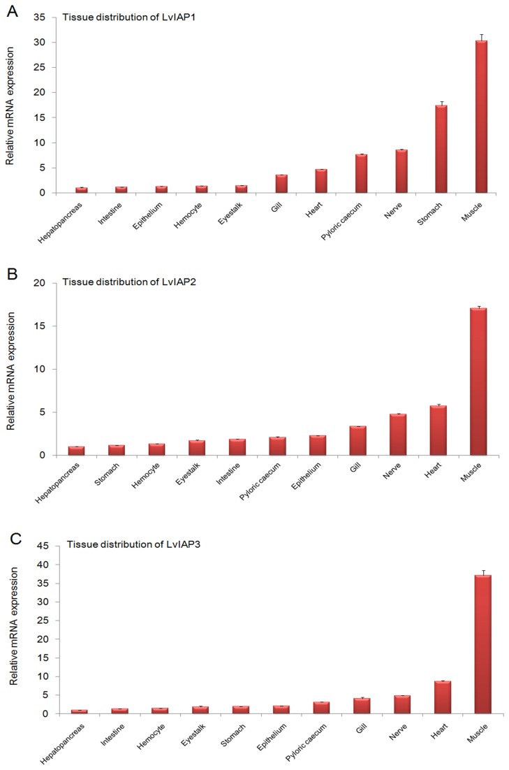
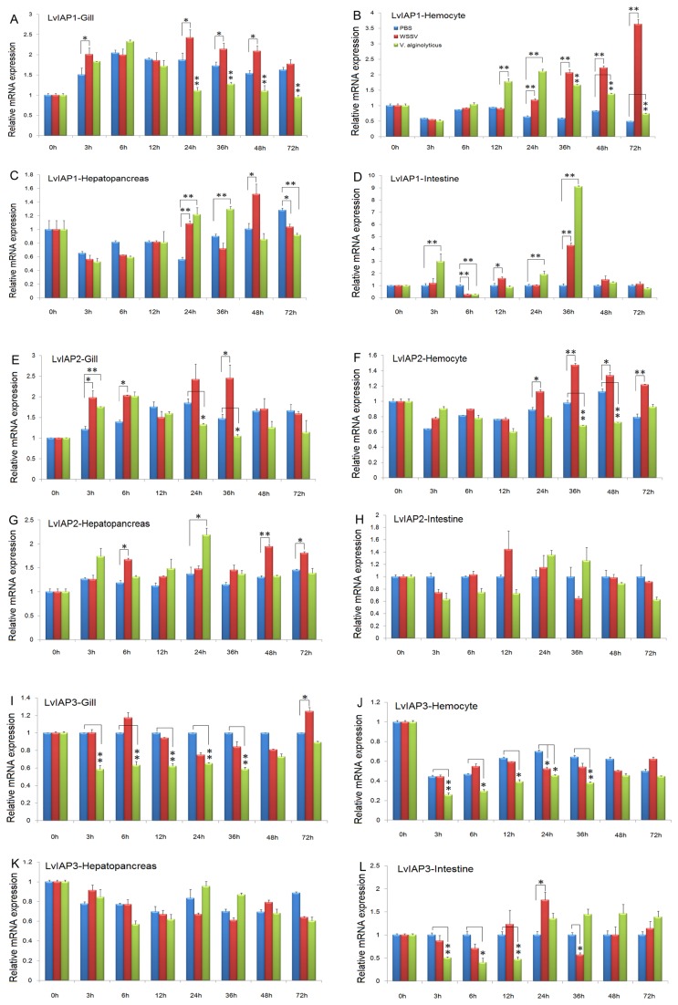
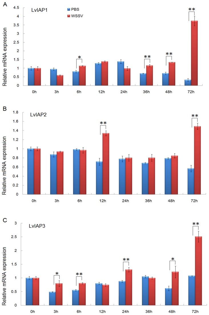
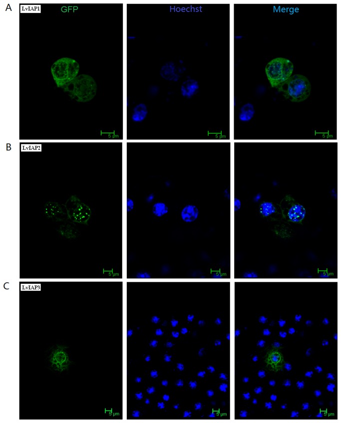
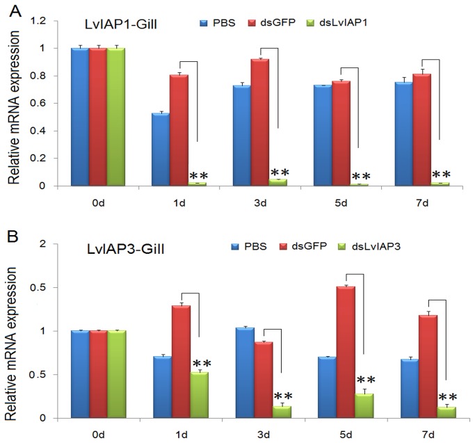
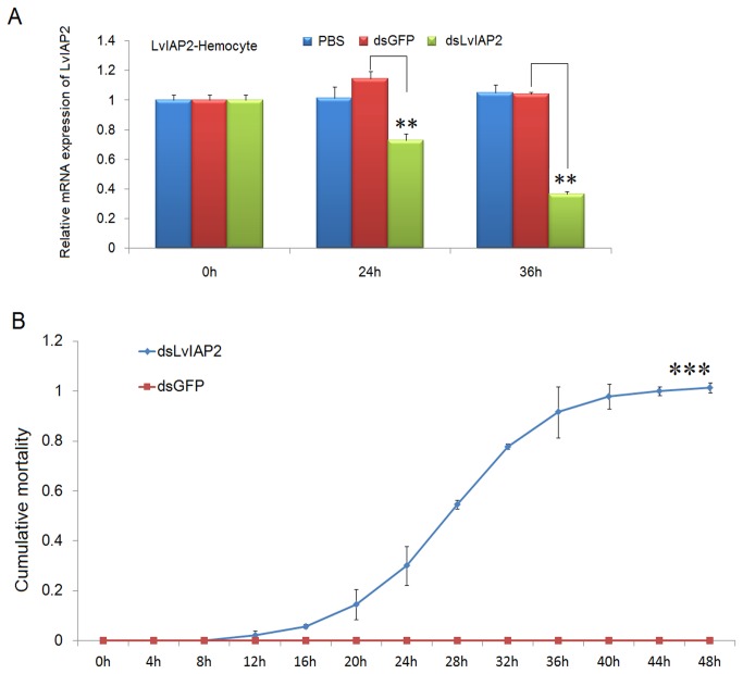
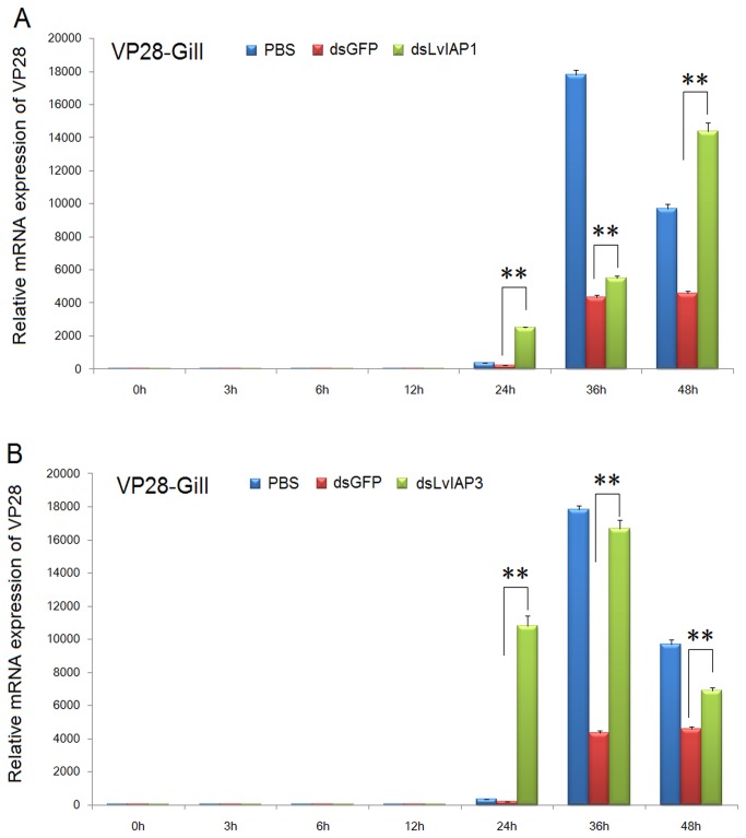
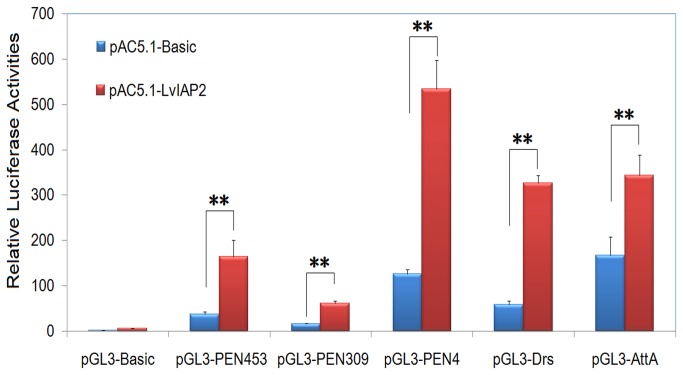
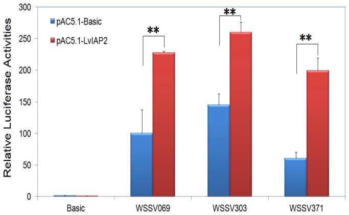
Inhibitors of apoptosis (IAPs) play important roles in apoptosis and NF-κB activation. In this study, we cloned and characterized three IAPs (LvIAP1-3) from the Pacific white shrimp, Litopenaeusvannamei. LvIAP1-3 proteins shared signature domains and exhibited significant similarities with other IAP family proteins. The tissue distributions of LvIAP1-3 were studied. The expression of LvIAP1-3 was induced in the muscle after white spot syndrome virus (WSSV) infection. LvIAP1 expression in the gill, hemocytes, hepatopancreas, and intestine was responsive to WSSV and Vibrioalginolyticus infections. LvIAP2 expression in the gill, hemocytes, and hepatopancreas was also responsive to WSSV infection. The expression of LvIAP3 in the gill, hemocytes, and intestine was reduced after V. alginolyticus infection. When overexpressed in Drosophila S2 cells, GFP labeled-LvIAP2 was distributed in the cytoplasm and appeared as speck-like aggregates in the nucleus. Both LvIAP1 and LvIAP3 were widely distributed throughout the cytoplasm and nucleus. The expression of LvIAP1, LvIAP2, and LvIAP3 was significantly knocked down by dsRNA-mediated gene silencing. In the gill of LvIAP1- or LvIAP3-silenced shrimp, the expression of WSSV VP28 was significantly higher than that of the dsGFP control group, suggesting that LvIAP1 and LvIAP3 may play protective roles in host defense against WSSV infection. Intriguingly, the LvIAP2-silenced shrimp all died within 48 hours after dsLvIAP2 injection. In the hemocytes of LvIAP2-silenced shrimps, the expression of antimicrobial peptide genes (AMPs), including Penaeidins, lysozyme, crustins, Vibriopenaeicidae-induced cysteine and proline-rich peptides (VICPs), was significantly downregulated, while the expression of anti-lipopolysaccharide factors (ALFs) was upregulated. Moreover, LvIAP2 activated the promoters of the NF-κB pathway-controlled AMPs, such as shrimp Penaeidins and Drosophila drosomycin and attacin A, in Drosophila S2 cells. Taken together, these results reveal that LvIAP1 and LvIAP3 might participate in the host defense against WSSV infection, and LvIAP2 might be involved in the regulation of shrimp AMPs.
凋亡抑制蛋白(IAPs)在凋亡和 NF-κB 激活中发挥重要作用。本研究从凡纳滨对虾(Litopenaeus vannamei)中克隆并鉴定了三种 IAP(LvIAP1-3)。LvIAP1-3 蛋白具有特征性结构域,与其他 IAP 家族蛋白具有显著相似性。研究了 LvIAP1-3 的组织分布。LvIAP1 在肌肉中被白斑综合征病毒(WSSV)感染诱导表达。LvIAP1 在虾的鳃、血细胞、肝胰腺和肠中对 WSSV 和溶藻弧菌感染有反应。LvIAP2 在虾的鳃、血细胞和肝胰腺中的表达也对 WSSV 感染有反应。LvIAP3 在虾的鳃、血细胞和肠中的表达在溶藻弧菌感染后减少。当在果蝇 S2 细胞中过表达时,GFP 标记的 LvIAP2 分布在细胞质中,并在核中呈现出斑点状聚集。LvIAP1 和 LvIAP3 都广泛分布于细胞质和核内。dsRNA 介导的基因沉默显著下调了 LvIAP1、LvIAP2 和 LvIAP3 的表达。在 LvIAP1 或 LvIAP3 沉默的虾的鳃中,WSSV VP28 的表达明显高于 dsGFP 对照组,表明 LvIAP1 和 LvIAP3 可能在宿主防御 WSSV 感染中发挥保护作用。有趣的是,LvIAP2 沉默的虾在注射 dsLvIAP2 后 48 小时内全部死亡。在 LvIAP2 沉默的虾血细胞中,抗菌肽基因(AMPs),包括 Penaeidins、溶菌酶、甲壳素、弧菌诱导的半胱氨酸和脯氨酸富含肽(VICPs)的表达显著下调,而抗脂多糖因子(ALFs)的表达上调。此外,LvIAP2 在果蝇 S2 细胞中激活了 NF-κB 通路控制的 AMPs 的启动子,如虾的 Penaeidins 和果蝇的 drosomycin 和 attacin A。综上所述,这些结果表明 LvIAP1 和 LvIAP3 可能参与宿主对 WSSV 感染的防御,LvIAP2 可能参与虾 AMPs 的调节。