Olin Neuropsychiatry Research Center, The Institute of Living/Hartford Hospital, 200 Retreat Ave, Whitehall Building, Hartford, CT, 06106, USA.
Brain Imaging Behav. 2014 Mar;8(1):73-86. doi: 10.1007/s11682-013-9246-z.
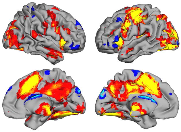
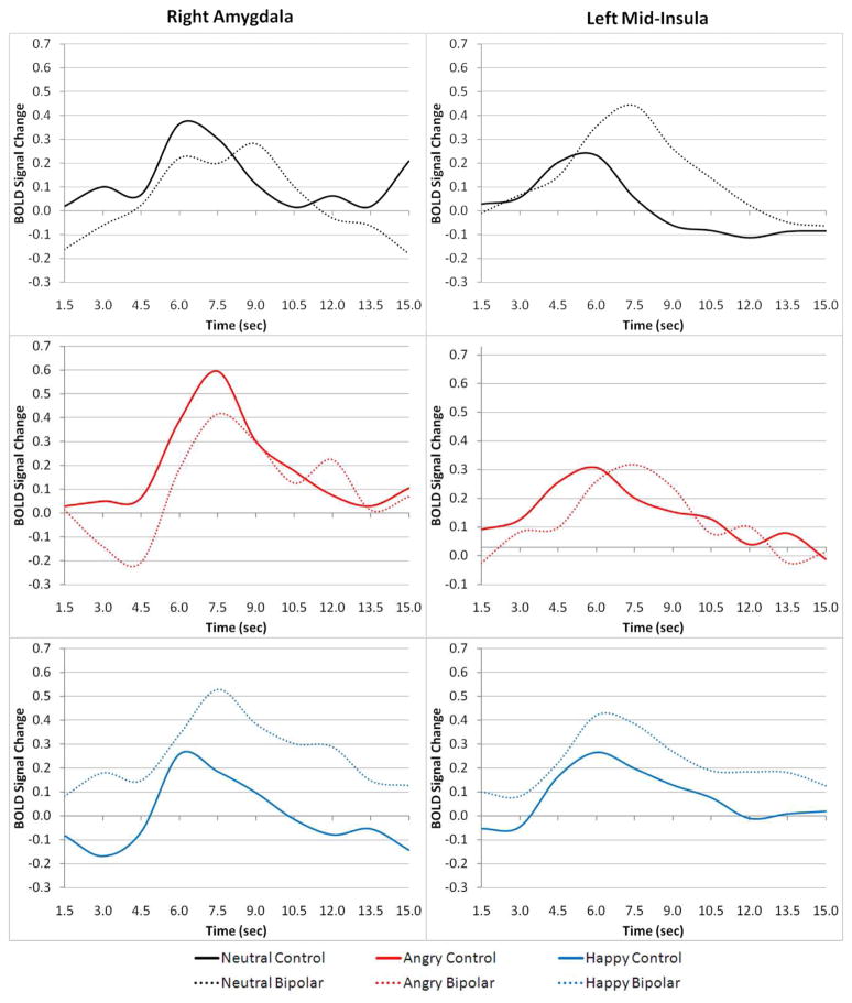
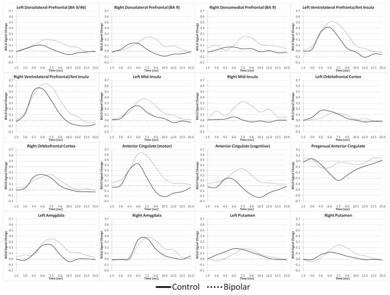
This fMRI study examined whether hemodynamic responses to affectively-salient stimuli were abnormally prolonged in remitted bipolar disorder, possibly representing a novel illness biomarker. A group of 18 DSM-IV bipolar I-diagnosed adults in remission and a demographically-matched control group performed an event-related fMRI gender-discrimination task in which face stimuli had task-irrelevant neutral, happy or angry expressions designed to elicit incidental emotional processing. Participants' brain activation was modeled using a "fully informed" SPM5 basis set. Mixed-model ANOVA tested for diagnostic group differences in BOLD response amplitude and shape within brain regions-of-interest selected from ALE meta-analysis of previous comparable fMRI studies. Bipolar-diagnosed patients had a generally longer duration and/or later-peaking hemodynamic response in amygdala and numerous prefrontal cortex brain regions. Data are consistent with existing models of bipolar limbic hyperactivity, but the prolonged frontolimbic response more precisely details abnormalities recognized in previous studies. Prolonged hemodynamic responses were unrelated to stimulus type, task performance, or degree of residual mood symptoms, suggesting an important novel trait vulnerability brain dysfunction in bipolar disorder. Bipolar patients also failed to engage pregenual cingulate and left orbitofrontal cortex-regions important to models of automatic emotion regulation-while engaging a delayed dorsolateral prefrontal cortex response not seen in controls. These results raise questions about whether there are meaningful relationships between bipolar dysfunction of specific ventromedial prefrontal cortex regions believed to automatically regulate emotional reactions and the prolonged responses in more lateral aspects of prefrontal cortex.
这项 fMRI 研究旨在探究在缓解期双相情感障碍中,情绪相关刺激的血液动力学反应是否异常延长,这可能代表一种新的疾病生物标志物。一组 18 名 DSM-IV 双相 I 型诊断缓解期患者和一组人口统计学匹配的对照组进行了一项事件相关 fMRI 性别识别任务,其中面孔刺激具有与任务无关的中性、快乐或愤怒表情,旨在引发偶然的情绪处理。使用“完全知情”的 SPM5 基础集来模拟参与者的大脑激活。混合模型方差分析测试了诊断组在大脑感兴趣区域的大脑活动强度和形状上的差异,这些区域是从先前类似 fMRI 研究的 ALE 荟萃分析中选择的。双相诊断患者的杏仁核和众多前额叶皮层脑区的血液动力学反应通常持续时间更长和/或峰值出现时间更晚。这些数据与双相情感边缘系统过度活跃的现有模型一致,但额叶边缘反应的延长更精确地详细说明了以前研究中识别出的异常。血液动力学反应与刺激类型、任务表现或残留情绪症状的严重程度无关,表明双相情感障碍中存在重要的新型特质易损性脑功能障碍。双相患者在参与自动情绪调节模型中重要的额下回扣带和左侧眶额皮层时未能参与,而在对照组中未观察到延迟的背外侧前额叶皮层反应。这些结果引发了关于是否存在特定腹内侧前额叶皮层区域的双相功能障碍与被认为自动调节情绪反应的更外侧前额叶皮层的延长反应之间存在有意义关系的问题。