Tu Ting-Yuan, Wang Zhe, Bai Jing, Sun Wei, Peng Weng Kung, Huang Ruby Yun-Ju, Thiery Jean-Paul, Kamm Roger D
BioSystems and Micromechanics IRG, Singapore-MIT Alliance for Research and Technology (SMART) Center, 1 CREATE Way, #04-13/14 Enterprise, Wing, Singapore, 138602, Singapore.
Adv Healthc Mater. 2014 Apr;3(4):609-16. doi: 10.1002/adhm.201300151. Epub 2013 Aug 27.
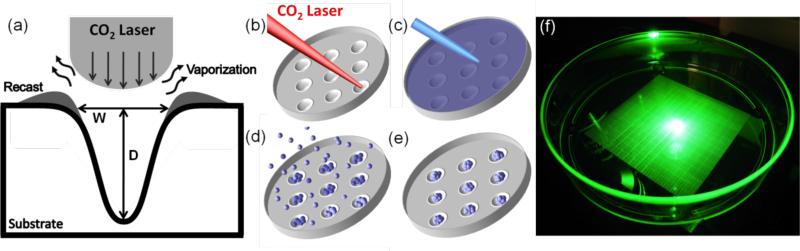
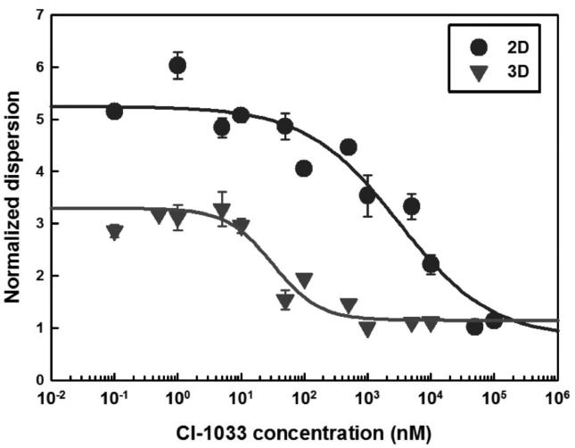
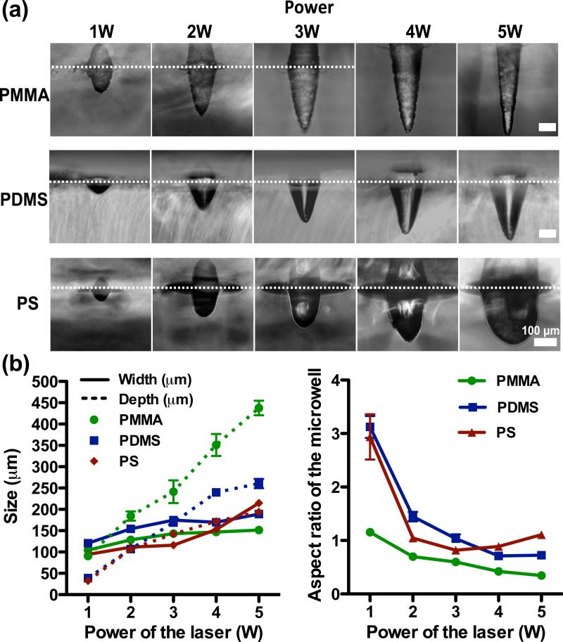
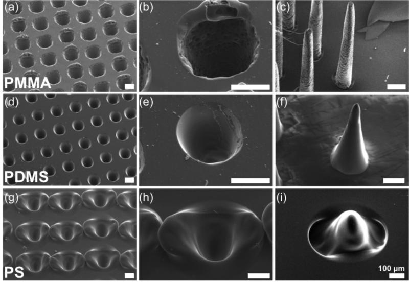
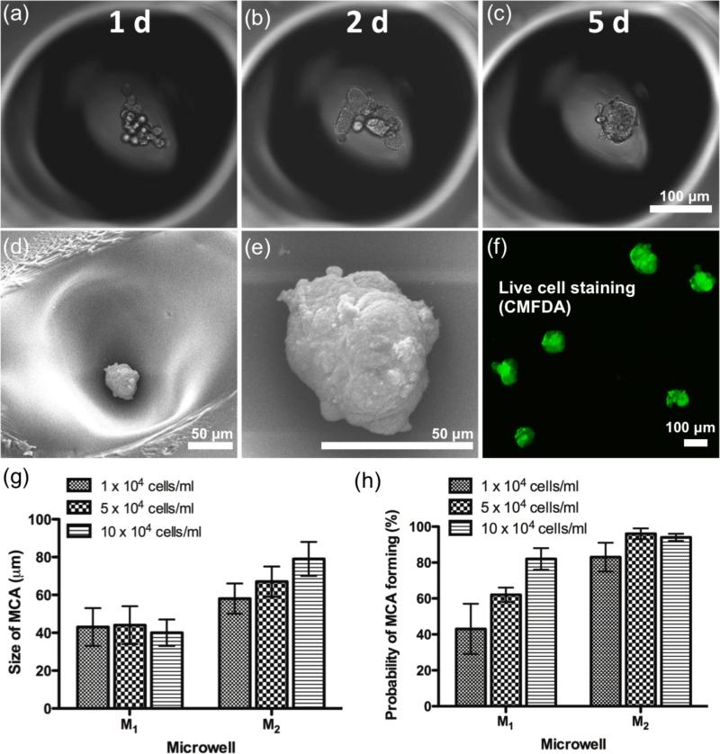
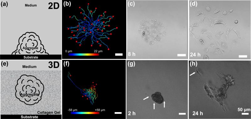
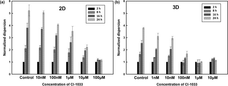
Microwell technology has revolutionized many aspects of in vitro cellular studies from 2D traditional cultures to 3D in vivo-like functional assays. However, existing lithography-based approaches are often costly and time-consuming. This study presents a rapid, low-cost prototyping method of CO2 laser ablation of a conventional untreated culture dish to create concave microwells used for generating multicellular aggregates, which can be readily available for general laboratories. Polymethylmethacrylate (PMMA), polydimethylsiloxane (PDMS), and polystyrene (PS) microwells are investigated, and each produces distinctive microwell features. Among these three materials, PS cell culture dishes produce the optimal surface smoothness and roundness. A549 lung cancer cells are grown to form cancer aggregates of controllable size from ≈40 to ≈80 μm in PS microwells. Functional assays of spheroids are performed to study migration on 2D substrates and in 3D hydrogel conditions as a step towards recapitulating the dissemination of cancer cells. Preclinical anti-cancer drug screening is investigated and reveals considerable differences between 2D and 3D conditions, indicating the importance of assay type as well as the utility of the present approach.
微孔技术已经彻底改变了体外细胞研究的许多方面,从二维传统培养到三维体内样功能分析。然而,现有的基于光刻的方法通常成本高昂且耗时。本研究提出了一种快速、低成本的原型制作方法,即通过二氧化碳激光烧蚀传统的未处理培养皿来创建用于生成多细胞聚集体的凹形微孔,这对于普通实验室来说很容易实现。对聚甲基丙烯酸甲酯(PMMA)、聚二甲基硅氧烷(PDMS)和聚苯乙烯(PS)微孔进行了研究,每种材料都产生了独特的微孔特征。在这三种材料中,PS细胞培养皿具有最佳的表面光滑度和圆度。在PS微孔中培养A549肺癌细胞,形成大小可控的癌聚集体,直径约为40至80μm。进行球体的功能分析,以研究在二维基质和三维水凝胶条件下的迁移,作为模拟癌细胞扩散的一步。研究了临床前抗癌药物筛选,结果显示二维和三维条件之间存在显著差异,这表明分析类型的重要性以及本方法的实用性。