• 文献检索
  • 文档翻译
  • 深度研究
  • 学术资讯
  • Suppr Zotero 插件Zotero 插件
  • 邀请有礼
  • 套餐&价格
  • 历史记录
应用&插件
Suppr Zotero 插件Zotero 插件浏览器插件Mac 客户端Windows 客户端微信小程序
定价
高级版会员购买积分包购买API积分包
服务
文献检索文档翻译深度研究API 文档MCP 服务
关于我们
关于 Suppr公司介绍联系我们用户协议隐私条款
关注我们

Suppr 超能文献

核心技术专利:CN118964589B侵权必究
粤ICP备2023148730 号-1Suppr @ 2026

文献检索

告别复杂PubMed语法,用中文像聊天一样搜索,搜遍4000万医学文献。AI智能推荐,让科研检索更轻松。

立即免费搜索

文件翻译

保留排版,准确专业,支持PDF/Word/PPT等文件格式,支持 12+语言互译。

免费翻译文档

深度研究

AI帮你快速写综述,25分钟生成高质量综述,智能提取关键信息,辅助科研写作。

立即免费体验

k-t GRAPPA加速的主动脉四维血流磁共振成像:对扫描时间、图像质量以及血流和壁面剪应力定量的影响

k-t GRAPPA accelerated four-dimensional flow MRI in the aorta: effect on scan time, image quality, and quantification of flow and wall shear stress.

作者信息

Schnell Susanne, Markl Michael, Entezari Pegah, Mahadewia Riti J, Semaan Edouard, Stankovic Zoran, Collins Jeremy, Carr James, Jung Bernd

机构信息

Department of Radiology, Northwestern University, Chicago, Illinois, USA.

出版信息

Magn Reson Med. 2014 Aug;72(2):522-33. doi: 10.1002/mrm.24925. Epub 2013 Sep 4.

DOI:10.1002/mrm.24925
PMID:24006309
原文链接:https://pmc.ncbi.nlm.nih.gov/articles/PMC4414256/
Abstract

PURPOSE

The purpose of this study was to evaluate the utility of k-t parallel imaging for accelerating aortic four-dimensional (4D)-flow MRI. The aim was to systematically investigate the impact of different acceleration factors and number of coil elements on acquisition time, image quality and quantification of hemodynamic parameters.

METHODS

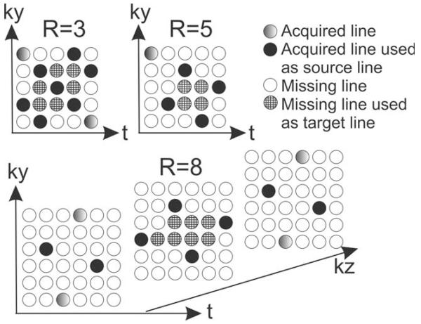
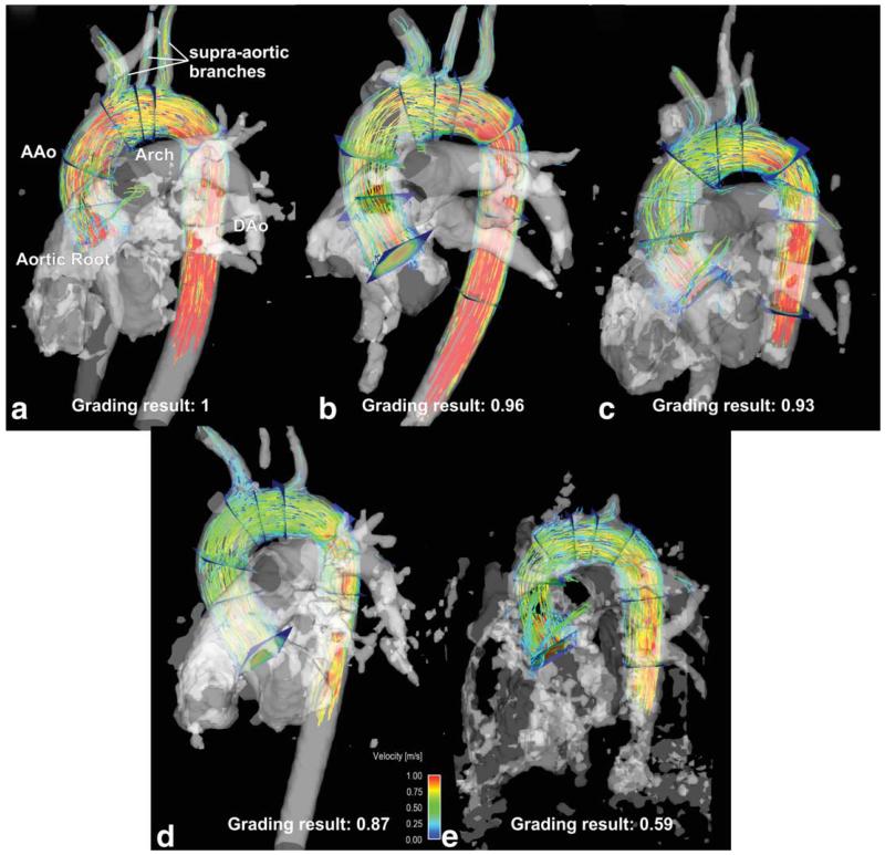
k-t accelerated 4D-flow MRI (spatial/temporal resolution = 2.1 × 2.5 × 2.5 mm/40.0 ms) was acquired in 10 healthy volunteers with acceleration factors R = 3, 5, and 8 using 12- and 32-channel receiver coils. Results were compared with conventional parallel imaging (GRAPPA [generalized autocalibrating partial parallel acquisition], R = 2). Data analysis included radiological grading of three-dimensional blood flow visualization quality as well as quantification of blood flow, velocities and wall shear stress (WSS).

RESULTS

k-t GRAPPA significantly reduced scan time by 28%, 54%, and 68%, for R = 3, 5, and 8, respectively, while maintaining image quality as demonstrated by overall similar image quality grading. Significant differences in peak WSS (diff12ch = -5.9%, diff32ch = 18.5%) and mean WSS (diff32ch = 13.9%) were found at the descending aorta for both receiver coils for R = 5 (PWSS < 0.04). Peak velocity differed for R=8 at the aortic root (-7.4%) and descending aorta (-12%) with PpeakVelo < 0.03.

CONCLUSION

k-t GRAPPA acceleration with a 12- or 32-channel receiver coil and an acceleration of 3 or 5 can compete with a standard GRAPPA R = 2 acceleration.

摘要

目的

本研究旨在评估k-t并行成像在加速主动脉四维(4D)血流磁共振成像(MRI)中的效用。目的是系统研究不同加速因子和线圈单元数量对采集时间、图像质量和血流动力学参数定量的影响。

方法

对10名健康志愿者采用12通道和32通道接收线圈,分别以加速因子R = 3、5和8进行k-t加速4D血流MRI(空间/时间分辨率 = 2.1×2.5×2.5 mm/40.0 ms)采集。将结果与传统并行成像(GRAPPA [广义自校准部分并行采集],R = 2)进行比较。数据分析包括对三维血流可视化质量进行放射学分级以及对血流、速度和壁面切应力(WSS)进行定量分析。

结果

对于R = 3、5和8,k-t GRAPPA分别显著减少扫描时间28%、54%和68%,同时通过总体相似的图像质量分级表明图像质量得以保持。对于R = 5,在两个接收线圈的降主动脉处均发现峰值WSS(12通道差异=-5.9%,32通道差异 = 18.5%)和平均WSS(32通道差异 = 13.9%)存在显著差异(峰值WSS < 0.04)。对于R = 8,在主动脉根部(-7.4%)和降主动脉(-12%)处峰值速度存在差异,峰值速度P < 0.03。

结论

采用12通道或32通道接收线圈且加速因子为3或5的k-t GRAPPA加速可与标准GRAPPA R = 2加速相媲美。

https://cdn.ncbi.nlm.nih.gov/pmc/blobs/5c96/4414256/a1f1d06082cb/nihms-683681-f0006.jpg
https://cdn.ncbi.nlm.nih.gov/pmc/blobs/5c96/4414256/be6aa6c3eae5/nihms-683681-f0001.jpg
https://cdn.ncbi.nlm.nih.gov/pmc/blobs/5c96/4414256/af7364e29e4f/nihms-683681-f0002.jpg
https://cdn.ncbi.nlm.nih.gov/pmc/blobs/5c96/4414256/51eb7b492be3/nihms-683681-f0003.jpg
https://cdn.ncbi.nlm.nih.gov/pmc/blobs/5c96/4414256/0b8fec482ee5/nihms-683681-f0004.jpg
https://cdn.ncbi.nlm.nih.gov/pmc/blobs/5c96/4414256/c9c627bc03ef/nihms-683681-f0005.jpg
https://cdn.ncbi.nlm.nih.gov/pmc/blobs/5c96/4414256/a1f1d06082cb/nihms-683681-f0006.jpg

相似文献

1
k-t GRAPPA accelerated four-dimensional flow MRI in the aorta: effect on scan time, image quality, and quantification of flow and wall shear stress.k-t GRAPPA加速的主动脉四维血流磁共振成像:对扫描时间、图像质量以及血流和壁面剪应力定量的影响
Magn Reson Med. 2014 Aug;72(2):522-33. doi: 10.1002/mrm.24925. Epub 2013 Sep 4.
2
K-t GRAPPA-accelerated 4D flow MRI of liver hemodynamics: influence of different acceleration factors on qualitative and quantitative assessment of blood flow.K-t GRAPPA加速的肝脏血流动力学4D流动MRI:不同加速因子对血流定性和定量评估的影响
MAGMA. 2015 Apr;28(2):149-59. doi: 10.1007/s10334-014-0456-1. Epub 2014 Aug 7.
3
Towards high-resolution 4D flow MRI in the human aorta using kt-GRAPPA and B1+ shimming at 7T.利用kt-GRAPPA和7T场强下的B1 +匀场技术实现人体主动脉高分辨率4D流动磁共振成像
J Magn Reson Imaging. 2016 Aug;44(2):486-99. doi: 10.1002/jmri.25164. Epub 2016 Feb 3.
4
Flow-sensitive 4D MRI of the thoracic aorta: comparison of image quality, quantitative flow, and wall parameters at 1.5 T and 3 T.胸主动脉的流量敏感 4D MRI:1.5T 和 3T 时图像质量、定量流量和管壁参数的比较。
J Magn Reson Imaging. 2012 Nov;36(5):1097-103. doi: 10.1002/jmri.23735. Epub 2012 Jun 28.
5
Accelerated dual-venc 4D flow MRI for neurovascular applications.用于神经血管应用的加速双静脉4D流动磁共振成像
J Magn Reson Imaging. 2017 Jul;46(1):102-114. doi: 10.1002/jmri.25595. Epub 2017 Feb 2.
6
k-t accelerated aortic 4D flow MRI in under two minutes: Feasibility and impact of resolution, k-space sampling patterns, and respiratory navigator gating on hemodynamic measurements.在两分钟内完成 k-t 加速主动脉 4D 流 MRI:分辨率、k 空间采样模式和呼吸导航门控对血流测量的可行性和影响。
Magn Reson Med. 2018 Jan;79(1):195-207. doi: 10.1002/mrm.26661. Epub 2017 Mar 7.
7
Quantification and comparison of 4D-flow MRI-derived wall shear stress and MRE-derived wall stiffness of the abdominal aorta.基于4D流磁共振成像的腹主动脉壁切应力与磁共振弹性成像的腹主动脉壁硬度的量化及比较
J Magn Reson Imaging. 2017 Mar;45(3):771-778. doi: 10.1002/jmri.25445. Epub 2016 Sep 7.
8
3.0T, time-resolved, 3D flow-sensitive MR in the thoracic aorta: Impact of k-t BLAST acceleration using 8- versus 32-channel coil arrays.3.0T时间分辨三维血流敏感磁共振成像在胸主动脉中的应用:8通道与32通道线圈阵列的k-t BLAST加速效果
J Magn Reson Imaging. 2015 Aug;42(2):495-504. doi: 10.1002/jmri.24814. Epub 2014 Dec 2.
9
Reproducibility and interobserver variability of systolic blood flow velocity and 3D wall shear stress derived from 4D flow MRI in the healthy aorta.健康主动脉中基于4D血流MRI得出的收缩期血流速度和三维壁面剪应力的可重复性及观察者间变异性
J Magn Reson Imaging. 2016 Jan;43(1):236-48. doi: 10.1002/jmri.24959. Epub 2015 Jul 3.
10
Thoracic aorta 3D hemodynamics in pediatric and young adult patients with bicuspid aortic valve.小儿及青年二叶式主动脉瓣患者的胸主动脉 3D 血流动力学。
J Magn Reson Imaging. 2015 Oct;42(4):954-63. doi: 10.1002/jmri.24847. Epub 2015 Jan 22.

引用本文的文献

1
Referenceless 4D flow cardiovascular magnetic resonance with deep learning.基于深度学习的无参考4D流心血管磁共振成像
J Cardiovasc Magn Reson. 2025 Jun 2;27(2):101920. doi: 10.1016/j.jocmr.2025.101920.
2
Highly accelerated 4D flow MRI with respiratory compensation and cardiac view sharing: a cross-sectional study of flow in the great vessels of pediatric congenital heart disease.具有呼吸补偿和心脏视图共享功能的高度加速4D流MRI:小儿先天性心脏病大血管血流的横断面研究。
Pediatr Radiol. 2025 May;55(6):1223-1234. doi: 10.1007/s00247-025-06226-1. Epub 2025 Apr 5.
3
Hemodynamic effects of entry and exit tear size in aortic dissection evaluated with in vitro magnetic resonance imaging and fluid-structure interaction simulation.

本文引用的文献

1
Aortic pulse wave velocity measurements with undersampled 4D flow-sensitive MRI: comparison with 2D and algorithm determination.应用欠采样 4D 流速敏感 MRI 测量主动脉脉搏波速度:与 2D 方法和算法测定的比较。
J Magn Reson Imaging. 2013 Apr;37(4):853-9. doi: 10.1002/jmri.23877. Epub 2012 Nov 2.
2
k-t BLAST and SENSE accelerated time-resolved three-dimensional phase contrast MRI in an intracranial aneurysm.颅内动脉瘤的 k-t BLAST 和 SENSE 加速时间分辨三维相位对比 MRI。
MAGMA. 2013 Jun;26(3):261-70. doi: 10.1007/s10334-012-0336-5. Epub 2012 Sep 7.
3
Evaluation of valvular insufficiency and shunts with parallel-imaging compressed-sensing 4D phase-contrast MR imaging with stereoscopic 3D velocity-fusion volume-rendered visualization.
体外磁共振成像和流固耦合模拟评估主动脉夹层入口和出口撕裂口大小对血流动力学的影响。
Sci Rep. 2023 Dec 18;13(1):22557. doi: 10.1038/s41598-023-49942-0.
4
4D Flow cardiovascular magnetic resonance consensus statement: 2023 update.4D Flow 心血管磁共振共识声明:2023 更新版。
J Cardiovasc Magn Reson. 2023 Jul 20;25(1):40. doi: 10.1186/s12968-023-00942-z.
5
Near-wall hemodynamic parameters quantification in in vitro intracranial aneurysms with 7 T PC-MRI.7T PC-MRI 检测颅内动脉瘤近壁血流动力学参数。
MAGMA. 2023 Apr;36(2):295-308. doi: 10.1007/s10334-023-01082-2. Epub 2023 Apr 19.
6
Accelerated sequences of 4D flow MRI using GRAPPA and compressed sensing: A comparison against conventional MRI and computational fluid dynamics.使用 GRAPPA 和压缩感知的 4D 流 MRI 加速序列:与常规 MRI 和计算流体动力学的比较。
Magn Reson Med. 2022 Dec;88(6):2432-2446. doi: 10.1002/mrm.29404. Epub 2022 Aug 25.
7
Accelerated dual-venc 4D flow MRI with variable high-venc spatial resolution for neurovascular applications.用于神经血管应用的可变高空间分辨率加速双门控 4D 流 MRI。
Magn Reson Med. 2022 Oct;88(4):1643-1658. doi: 10.1002/mrm.29306. Epub 2022 Jun 26.
8
FlowRAU-Net: Accelerated 4D Flow MRI of Aortic Valvular Flows With a Deep 2D Residual Attention Network.FlowRAU-Net:基于深度二维残差注意力网络的主动脉瓣血流加速 4D Flow MRI
IEEE Trans Biomed Eng. 2022 Dec;69(12):3812-3824. doi: 10.1109/TBME.2022.3180691. Epub 2022 Nov 23.
9
Technical Background for 4D Flow MR Imaging.4D 流磁共振成像的技术背景。
Magn Reson Med Sci. 2022 Mar 1;21(2):267-277. doi: 10.2463/mrms.rev.2021-0104. Epub 2022 Feb 11.
10
The role of 4D flow MRI for clinical applications in cardiovascular disease: current status and future perspectives.4D 流动磁共振成像在心血管疾病临床应用中的作用:现状与未来展望。
Quant Imaging Med Surg. 2021 Sep;11(9):4193-4210. doi: 10.21037/qims-20-1234.
采用立体 3D 速度融合容积渲染可视化的平行成像压缩感知 4D 相位对比 MR 成像评估瓣膜功能不全和分流。
Radiology. 2012 Oct;265(1):87-95. doi: 10.1148/radiol.12120055. Epub 2012 Aug 24.
4
Highly undersampled phase-contrast flow measurements using compartment-based k-t principal component analysis.基于分室的 k-t 主成分分析的高欠采样相位对比流量测量。
Magn Reson Med. 2013 Feb;69(2):434-43. doi: 10.1002/mrm.24273. Epub 2012 Apr 23.
5
Rapid pediatric cardiac assessment of flow and ventricular volume with compressed sensing parallel imaging volumetric cine phase-contrast MRI.应用压缩感知并行成像容积电影相位对比 MRI 快速儿科心脏心排量和心室容量评估。
AJR Am J Roentgenol. 2012 Mar;198(3):W250-9. doi: 10.2214/AJR.11.6969.
6
4D cardiovascular magnetic resonance velocity mapping of alterations of right heart flow patterns and main pulmonary artery hemodynamics in tetralogy of Fallot.四叶式法洛四联症右心血流模式和主肺动脉血流动力学改变的 4D 心血管磁共振速度成像。
J Cardiovasc Magn Reson. 2012 Feb 7;14(1):16. doi: 10.1186/1532-429X-14-16.
7
Four-dimensional phase contrast MRI with accelerated dual velocity encoding.四维相位对比磁共振成像加速双流速编码技术。
J Magn Reson Imaging. 2012 Jun;35(6):1462-71. doi: 10.1002/jmri.23588. Epub 2012 Jan 26.
8
Four-dimensional flow-sensitive MRI of the thoracic aorta: 12- versus 32-channel coil arrays.胸主动脉的四维血流敏感 MRI:12 通道与 32 通道线圈阵列。
J Magn Reson Imaging. 2012 Jan;35(1):190-5. doi: 10.1002/jmri.22633. Epub 2011 Oct 11.
9
Quantification and visualization of cardiovascular 4D velocity mapping accelerated with parallel imaging or k-t BLAST: head to head comparison and validation at 1.5 T and 3 T.心血管四维速度图的定量和可视化加速并行成像或 k-t BLAST:1.5T 和 3T 的头对头比较和验证。
J Cardiovasc Magn Reson. 2011 Oct 4;13(1):55. doi: 10.1186/1532-429X-13-55.
10
Reproducibility of flow and wall shear stress analysis using flow-sensitive four-dimensional MRI.采用血流敏感四维 MRI 分析血流和壁切应力的可重复性。
J Magn Reson Imaging. 2011 Apr;33(4):988-94. doi: 10.1002/jmri.22519.