Department of Neuroscience and Imaging, University "G. d'Annunzio" Chieti-Pescara, Chieti, Italy.
Molecular Neurology Unit, Center of Excellence on Aging, University "G. d'Annunzio", Chieti-Pescara, Chieti, Italy.
PeerJ. 2013 Aug 20;1:e135. doi: 10.7717/peerj.135. eCollection 2013.
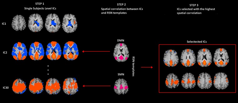
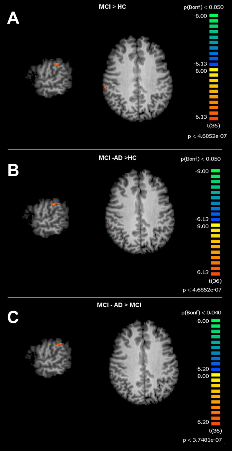
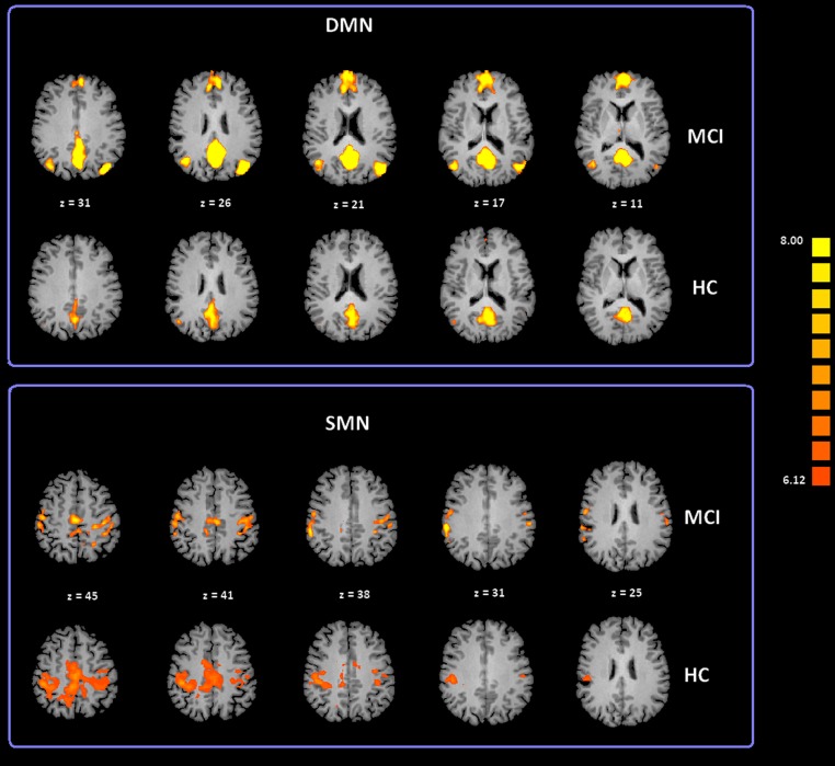
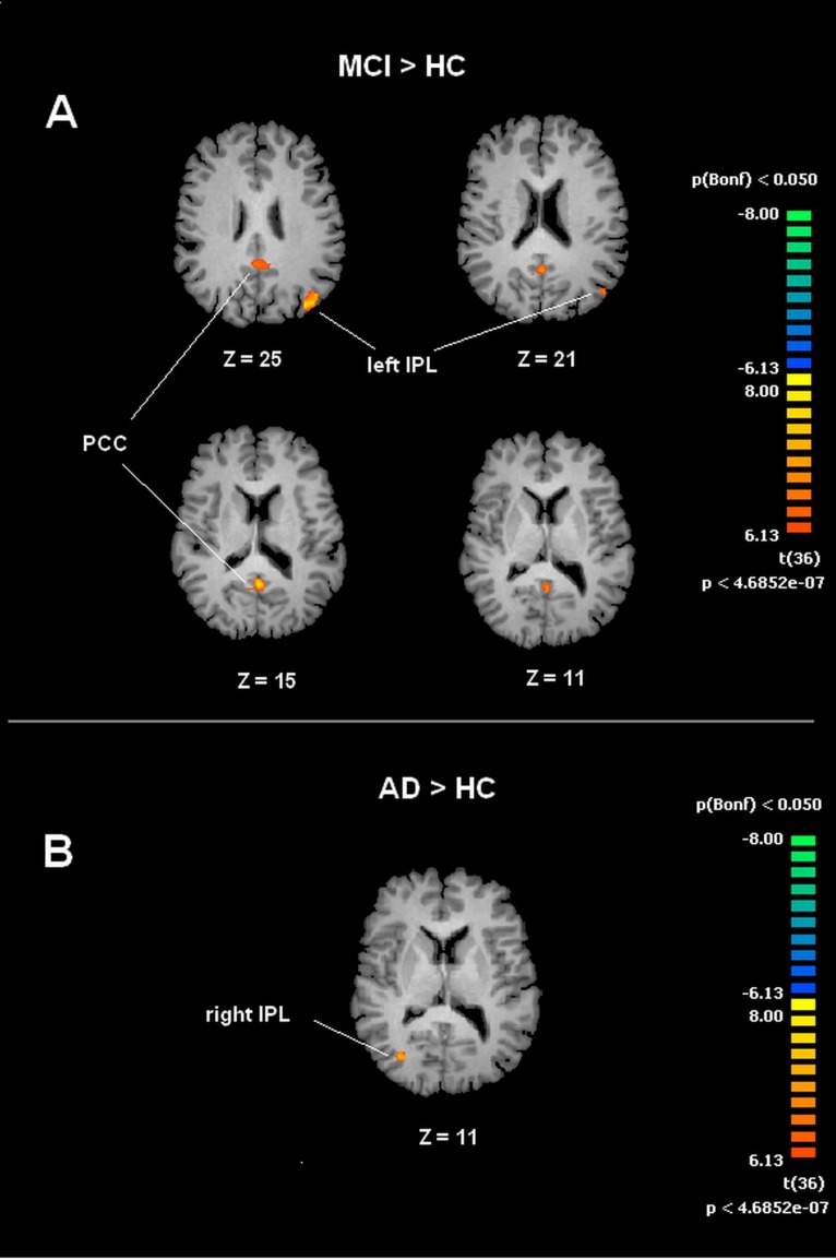
Objectives. Aging is the major risk factor for Alzheimer Disease (AD) and Mild Cognitive Impairment (MCI). The aim of this study was to identify novel modifications of brain functional connectivity in MCI patients. MCI individuals were compared to healthy elderly subjects. Methods. We enrolled 37 subjects (age range 60-80 y.o.). Of these, 13 subjects were affected by MCI and 24 were age-matched healthy elderly control (HC). Subjects were evaluated with Mini Mental State Examination (MMSE), Frontal Assessment Battery (FAB), and prose memory (Babcock story) tests. In addition, with functional Magnetic Resonance Imaging (fMRI), we investigated resting state network (RSN) activities. Resting state (Rs) fMRI data were analyzed by means of Independent Component Analysis (ICA). Subjects were followed-up with neuropsychological evaluations for three years. Results. Rs-fMRI of MCI subjects showed increased intrinsic connectivity in the Default Mode Network (DMN) and in the Somatomotor Network (SMN). Analysis of the DMN showed statistically significant increased activation in the posterior cingulate cortex (PCC) and left inferior parietal lobule (lIPL). During the three years follow-up, 4 MCI subjects converted to AD. The subset of MCI AD-converted patients showed increased connectivity in the right Inferior Parietal Lobule (rIPL). As for SMN activity, MCI and MCI-AD converted groups showed increased level of connectivity in correspondence of the right Supramarginal Gyrus (rSG). Conclusions. Our findings indicate alterations of DMN and SMN activity in MCI subjects, thereby providing potential imaging-based markers that can be helpful for the early diagnosis and monitoring of these patients.
目的。衰老(aging)是阿尔茨海默病(Alzheimer Disease,AD)和轻度认知障碍(Mild Cognitive Impairment,MCI)的主要危险因素。本研究旨在鉴定 MCI 患者大脑功能连接的新修饰。将 MCI 患者与健康老年对照(healthy elderly subjects)进行比较。
方法。我们纳入了 37 名受试者(年龄 60-80 岁)。其中,13 名受试者患有 MCI,24 名受试者为年龄匹配的健康老年对照(HC)。受试者接受了简易精神状态检查(Mini Mental State Examination,MMSE)、额叶评估量表(Frontal Assessment Battery,FAB)和散文记忆(Babcock 故事)测试。此外,我们通过功能磁共振成像(functional Magnetic Resonance Imaging,fMRI)来研究静息状态网络(resting state network,RSN)活动。静息状态(resting state,Rs)fMRI 数据通过独立成分分析(Independent Component Analysis,ICA)进行分析。对受试者进行了为期三年的神经心理学随访。
结果。MCI 受试者的 Rs-fMRI 显示默认模式网络(Default Mode Network,DMN)和躯体运动网络(Somatomotor Network,SMN)的固有连接增加。DMN 的分析显示后扣带回皮层(posterior cingulate cortex,PCC)和左侧顶下小叶(left inferior parietal lobule,lIPL)的激活显著增加。在三年的随访中,4 名 MCI 患者转化为 AD。MCI-AD 转化患者亚组显示右侧顶下小叶(right inferior parietal lobule,rIPL)的连接增加。对于 SMN 活动,MCI 和 MCI-AD 转化组在右侧缘上回(right supramarginal gyrus,rSG)处显示出更高的连接水平。
结论。我们的研究结果表明 MCI 患者的 DMN 和 SMN 活动发生改变,从而提供了潜在的基于影像学的标志物,有助于对这些患者进行早期诊断和监测。