Hou Yanbing, Yang Jing, Luo Chunyan, Song Wei, Ou Ruwei, Liu Wanglin, Gong Qiyong, Shang Huifang
Department of Neurology, West China Hospital, Sichuan University Chengdu, China.
Huaxi MR Research Center, Department of Radiology, West China Hospital, Sichuan University Chengdu, China.
Front Aging Neurosci. 2016 Oct 26;8:247. doi: 10.3389/fnagi.2016.00247. eCollection 2016.
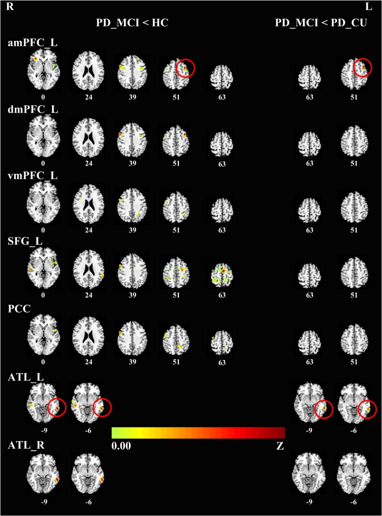
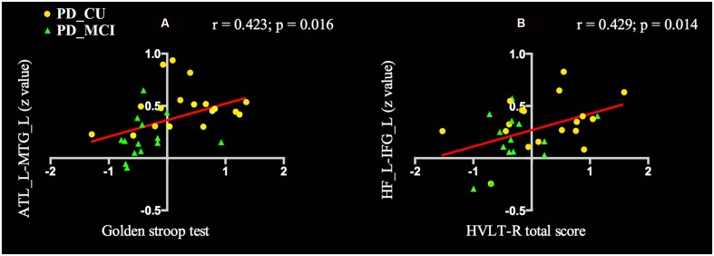
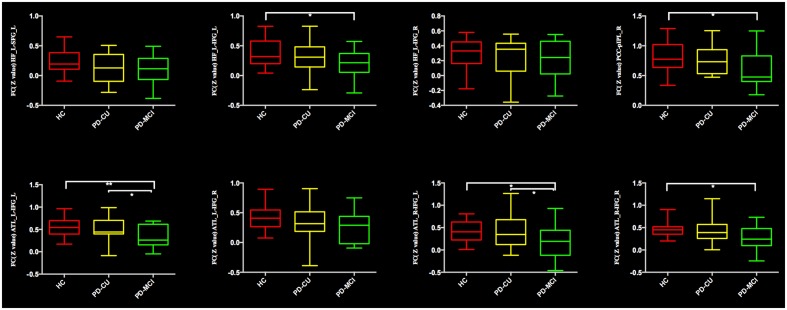
Cognitive impairments are common in Parkinson's disease (PD) and can even occur in the early stages. The default mode network (DMN) is highly relevant for cognitive processes; however, it remains largely unknown if changes in the DMN connectivity are related to the cognitive decline in drug-naïve early stage PD patients with a mild cognitive impairment (MCI). This study used resting-state functional MRI (fMRI) to explore the brain connectivity of the DMN in early stage drug-naïve PD patients with MCI. We recruited 32 early stage drug-naïve PD patients and 22 matched healthy controls (HC). Among the PD patients, 14 were classified as having MCI (PD-MCI) and 18 were classified as having unimpaired cognition (PD-CU). The functional integration of the DMN was evaluated by a seed-based correlation approach. The brain connectivity analysis revealed reduced functional connectivity (FC) in both PD subgroups compared with HC. The PD-MCI group showed a significant reduction in FC between the DMN and a set of regions, including the precentral gyrus, middle temporal gyrus, insula, anterior inferior parietal lobule and middle frontal gyrus. Compared to the PD-CU group, the PD-MCI group demonstrated a significantly decreased FC in the middle frontal and middle temporal gyri. Additionally, compared to HC, the PD-MCI group had a significantly decreased FC within the DMN, mainly in the FC between the hippocampal formation and inferior frontal gyrus, between the posterior cingulate cortex and posterior inferior parietal lobule, and between the anterior temporal lobe and inferior frontal gyrus. Compared to the PD-CU group, the only significantly decreased FC within the DMN in the PD-MCI group was between the anterior temporal lobe and inferior frontal gyrus. In all PD patients, the decreased FC between anterior temporal lobe and middle temporal gyrus was positively correlated with attention/working performance, and the reduced FC between the hippocampal formation and inferior frontal gyrus was also positively correlated with memory function. Our findings suggest that an altered DMN connectivity characterizes PD-MCI patients. These findings may be helpful for facilitating the further understanding of the potential mechanisms underlying MCI in PD. However, our results are preliminary, and further investigation is needed.
认知障碍在帕金森病(PD)中很常见,甚至在疾病早期就可能出现。默认模式网络(DMN)与认知过程高度相关;然而,对于未经药物治疗的早期轻度认知障碍(MCI)帕金森病患者,DMN连接性的变化是否与认知功能下降有关,目前仍知之甚少。本研究采用静息态功能磁共振成像(fMRI)来探究未经药物治疗的早期MCI帕金森病患者的DMN脑连接性。我们招募了32例未经药物治疗的早期帕金森病患者和22例匹配的健康对照(HC)。在帕金森病患者中,14例被归类为患有MCI(PD-MCI),18例被归类为认知功能未受损(PD-CU)。通过基于种子点的相关性方法评估DMN的功能整合。脑连接性分析显示,与健康对照相比,两个帕金森病亚组的功能连接(FC)均降低。PD-MCI组在DMN与一组区域之间的FC显著降低,这些区域包括中央前回、颞中回、岛叶、顶下小叶前部和额中回。与PD-CU组相比,PD-MCI组在额中回和颞中回的FC显著降低。此外,与健康对照相比,PD-MCI组在DMN内的FC显著降低,主要是海马结构与额下回之间、后扣带回皮质与顶下小叶后部之间以及颞叶前部与额下回之间的FC。与PD-CU组相比,PD-MCI组在DMN内唯一显著降低的FC是颞叶前部与额下回之间。在所有帕金森病患者中,颞叶前部与颞中回之间FC的降低与注意力/工作表现呈正相关,海马结构与额下回之间FC的降低也与记忆功能呈正相关。我们的研究结果表明,DMN连接性改变是PD-MCI患者的特征。这些发现可能有助于进一步理解帕金森病中MCI的潜在机制。然而,我们的结果是初步的,需要进一步研究。