• 文献检索
  • 文档翻译
  • 深度研究
  • 学术资讯
  • Suppr Zotero 插件Zotero 插件
  • 邀请有礼
  • 套餐&价格
  • 历史记录
应用&插件
Suppr Zotero 插件Zotero 插件浏览器插件Mac 客户端Windows 客户端微信小程序
定价
高级版会员购买积分包购买API积分包
服务
文献检索文档翻译深度研究API 文档MCP 服务
关于我们
关于 Suppr公司介绍联系我们用户协议隐私条款
关注我们

Suppr 超能文献

核心技术专利:CN118964589B侵权必究
粤ICP备2023148730 号-1Suppr @ 2026

文献检索

告别复杂PubMed语法,用中文像聊天一样搜索,搜遍4000万医学文献。AI智能推荐,让科研检索更轻松。

立即免费搜索

文件翻译

保留排版,准确专业,支持PDF/Word/PPT等文件格式,支持 12+语言互译。

免费翻译文档

深度研究

AI帮你快速写综述,25分钟生成高质量综述,智能提取关键信息,辅助科研写作。

立即免费体验

自动密度:一种测量乳腺钼靶密度的自动化方法,可预测乳腺癌风险和筛查结果。

AutoDensity: an automated method to measure mammographic breast density that predicts breast cancer risk and screening outcomes.

作者信息

Nickson Carolyn, Arzhaeva Yulia, Aitken Zoe, Elgindy Tarek, Buckley Mitchell, Li Min, English Dallas R, Kavanagh Anne M

出版信息

Breast Cancer Res. 2013;15(5):R80. doi: 10.1186/bcr3474.

DOI:10.1186/bcr3474
PMID:24020331
原文链接:https://pmc.ncbi.nlm.nih.gov/articles/PMC3978575/
Abstract

INTRODUCTION

While Cumulus - a semi-automated method for measuring breast density - is utilised extensively in research, it is labour-intensive and unsuitable for screening programmes that require an efficient and valid measure on which to base screening recommendations. We develop an automated method to measure breast density (AutoDensity) and compare it to Cumulus in terms of association with breast cancer risk and breast cancer screening outcomes.

METHODS

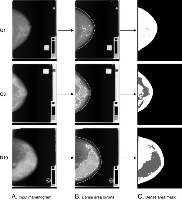
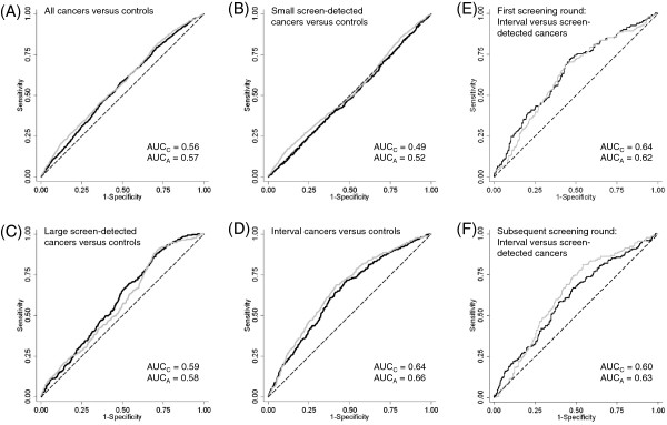
AutoDensity automatically identifies the breast area in the mammogram and classifies breast density in a similar way to Cumulus, through a fast, stand-alone Windows or Linux program. Our sample comprised 985 women with screen-detected cancers, 367 women with interval cancers and 4,975 controls (women who did not have cancer), sampled from first and subsequent screening rounds of a film mammography screening programme. To test the validity of AutoDensity, we compared the effect estimates using AutoDensity with those using Cumulus from logistic regression models that tested the association between breast density and breast cancer risk, risk of small and large screen-detected cancers and interval cancers, and screening programme sensitivity (the proportion of cancers that are screen-detected). As a secondary analysis, we report on correlation between AutoDensity and Cumulus measures.

RESULTS

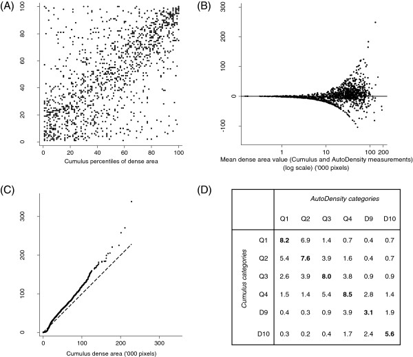
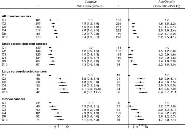
AutoDensity performed similarly to Cumulus in all associations tested. For example, using AutoDensity, the odds ratios for women in the highest decile of breast density compared to women in the lowest quintile for invasive breast cancer, interval cancers, large and small screen-detected cancers were 3.2 (95% CI 2.5 to 4.1), 4.7 (95% CI 3.0 to 7.4), 6.4 (95% CI 3.7 to 11.1) and 2.2 (95% CI 1.6 to 3.0) respectively. For Cumulus the corresponding odds ratios were: 2.4 (95% CI 1.9 to 3.1), 4.1 (95% CI 2.6 to 6.3), 6.6 (95% CI 3.7 to 11.7) and 1.3 (95% CI 0.9 to 1.8). Correlation between Cumulus and AutoDensity measures was 0.63 (P < 0.001).

CONCLUSIONS

Based on the similarity of the effect estimates for AutoDensity and Cumulus inmodels of breast density and breast cancer risk and screening outcomes, we conclude that AutoDensity is a valid automated method for measuring breast density from digitised film mammograms.

摘要

引言

虽然积云法(Cumulus)——一种测量乳房密度的半自动方法——在研究中被广泛使用,但它劳动强度大,不适用于需要高效且有效测量方法以制定筛查建议的筛查项目。我们开发了一种自动测量乳房密度的方法(自动密度法,AutoDensity),并在与乳腺癌风险及乳腺癌筛查结果的关联方面将其与积云法进行比较。

方法

自动密度法通过一个快速的独立Windows或Linux程序,自动识别乳腺钼靶片中的乳房区域,并以与积云法类似的方式对乳房密度进行分类。我们的样本包括985名经筛查发现患有癌症的女性、367名患有间期癌的女性以及4975名对照者(未患癌症的女性),这些样本取自一个乳腺钼靶筛查项目的首次及后续筛查轮次。为了测试自动密度法的有效性,我们将使用自动密度法得到的效应估计值与使用积云法从逻辑回归模型中得到的效应估计值进行比较,这些逻辑回归模型测试了乳房密度与乳腺癌风险、大小不同的经筛查发现的癌症及间期癌的风险以及筛查项目敏感性(经筛查发现的癌症比例)之间的关联。作为一项次要分析,我们报告了自动密度法与积云法测量值之间的相关性。

结果

在所有测试的关联中,自动密度法的表现与积云法相似。例如,使用自动密度法时,与乳房密度最低五分位数的女性相比,乳房密度最高十分位数的女性患浸润性乳腺癌、间期癌、大小不同的经筛查发现的癌症的优势比分别为3.2(95%可信区间2.5至4.1)、4.7(95%可信区间3.0至7.4)、6.4(95%可信区间3.7至11.1)和2.2(95%可信区间1.6至3.0)。对于积云法,相应的优势比分别为:2.4(95%可信区间1.9至3.1)、4.1(95%可信区间2.6至6.3)、6.6(95%可信区间3.7至11.7)和1.3(95%可信区间0.9至1.8)。积云法与自动密度法测量值之间的相关性为0.63(P<0.001)。

结论

基于自动密度法和积云法在乳房密度及乳腺癌风险和筛查结果模型中的效应估计值的相似性,我们得出结论,自动密度法是一种从数字化乳腺钼靶片中测量乳房密度的有效自动化方法。

https://cdn.ncbi.nlm.nih.gov/pmc/blobs/92aa/3978575/228c35175406/bcr3474-5.jpg
https://cdn.ncbi.nlm.nih.gov/pmc/blobs/92aa/3978575/1a080a37f62e/bcr3474-1.jpg
https://cdn.ncbi.nlm.nih.gov/pmc/blobs/92aa/3978575/b8fad03f7297/bcr3474-2.jpg
https://cdn.ncbi.nlm.nih.gov/pmc/blobs/92aa/3978575/cb9d7cb5f44f/bcr3474-3.jpg
https://cdn.ncbi.nlm.nih.gov/pmc/blobs/92aa/3978575/ce47ee950ca9/bcr3474-4.jpg
https://cdn.ncbi.nlm.nih.gov/pmc/blobs/92aa/3978575/228c35175406/bcr3474-5.jpg

相似文献

1
AutoDensity: an automated method to measure mammographic breast density that predicts breast cancer risk and screening outcomes.自动密度:一种测量乳腺钼靶密度的自动化方法,可预测乳腺癌风险和筛查结果。
Breast Cancer Res. 2013;15(5):R80. doi: 10.1186/bcr3474.
2
A comparison of five methods of measuring mammographic density: a case-control study.五种测量乳腺密度方法的比较:病例对照研究。
Breast Cancer Res. 2018 Feb 5;20(1):10. doi: 10.1186/s13058-018-0932-z.
3
Mammographic density defined by higher than conventional brightness threshold better predicts breast cancer risk for full-field digital mammograms.通过高于传统亮度阈值定义的乳腺X线摄影密度能更好地预测全视野数字化乳腺X线摄影的乳腺癌风险。
Breast Cancer Res. 2015 Nov 18;17:142. doi: 10.1186/s13058-015-0654-4.
4
Predicting interval and screen-detected breast cancers from mammographic density defined by different brightness thresholds.根据不同亮度阈值定义的乳腺密度预测间期和筛查性乳腺癌。
Breast Cancer Res. 2018 Dec 13;20(1):152. doi: 10.1186/s13058-018-1081-0.
5
The influence of mammogram acquisition on the mammographic density and breast cancer association in the Mayo Mammography Health Study cohort.在梅奥乳腺X线摄影健康研究队列中,乳腺X线摄影采集对乳腺X线密度和乳腺癌关联的影响。
Breast Cancer Res. 2012 Nov 15;14(6):R147. doi: 10.1186/bcr3357.
6
Risk factors and tumor characteristics of interval cancers by mammographic density.基于乳腺密度的间隔期癌症的风险因素和肿瘤特征。
J Clin Oncol. 2015 Mar 20;33(9):1030-7. doi: 10.1200/JCO.2014.58.9986. Epub 2015 Feb 2.
7
Comparison of fully and semi-automated area-based methods for measuring mammographic density and predicting breast cancer risk.基于面积的全自动和半自动方法测量乳腺密度和预测乳腺癌风险的比较。
Br J Cancer. 2014 Apr 2;110(7):1908-16. doi: 10.1038/bjc.2014.82. Epub 2014 Feb 20.
8
Mammographic features associated with interval breast cancers in screening programs.筛查项目中与间期乳腺癌相关的乳腺钼靶特征。
Breast Cancer Res. 2014 Aug 26;16(4):417. doi: 10.1186/s13058-014-0417-7.
9
High-throughput mammographic-density measurement: a tool for risk prediction of breast cancer.高通量乳腺密度测量:一种乳腺癌风险预测工具。
Breast Cancer Res. 2012 Jul 30;14(4):R114. doi: 10.1186/bcr3238.
10
Digital versus screen-film mammography: impact of mammographic density and hormone therapy on breast cancer detection.数字化乳腺钼靶摄影与屏-片乳腺钼靶摄影:乳腺密度和激素治疗对乳腺癌检测的影响
Breast Cancer Res Treat. 2015 Nov;154(2):377-87. doi: 10.1007/s10549-015-3622-x. Epub 2015 Oct 30.

引用本文的文献

1
Cohort Profile: Lifepool.队列简介:生命池。
Int J Epidemiol. 2025 Apr 12;54(3). doi: 10.1093/ije/dyaf044.
2
Sentinel Lymph Node Biopsy in Breast Cancer Using Different Types of Tracers According to Molecular Subtypes and Breast Density-A Randomized Clinical Study.根据分子亚型和乳腺密度使用不同类型示踪剂的乳腺癌前哨淋巴结活检——一项随机临床研究
Diagnostics (Basel). 2024 Oct 31;14(21):2439. doi: 10.3390/diagnostics14212439.
3
Breast Cancer Risk Assessment: A Review on Mammography-Based Approaches.乳腺癌风险评估:基于乳腺X线摄影方法的综述

本文引用的文献

1
High-throughput mammographic-density measurement: a tool for risk prediction of breast cancer.高通量乳腺密度测量:一种乳腺癌风险预测工具。
Breast Cancer Res. 2012 Jul 30;14(4):R114. doi: 10.1186/bcr3238.
2
A novel automated mammographic density measure and breast cancer risk.一种新型的自动乳腺密度测量方法与乳腺癌风险。
J Natl Cancer Inst. 2012 Jul 3;104(13):1028-37. doi: 10.1093/jnci/djs254.
3
Detection of breast cancer with addition of annual screening ultrasound or a single screening MRI to mammography in women with elevated breast cancer risk.
J Imaging. 2021 Jun 12;7(6):98. doi: 10.3390/jimaging7060098.
4
The cost-effectiveness of risk-stratified breast cancer screening in the UK.英国风险分层乳腺癌筛查的成本效益。
Br J Cancer. 2023 Nov;129(11):1801-1809. doi: 10.1038/s41416-023-02461-1. Epub 2023 Oct 17.
5
Beyond Breast Density: Risk Measures for Breast Cancer in Multiple Imaging Modalities.超越乳腺密度:多种影像学模式下乳腺癌的风险评估指标。
Radiology. 2023 Mar;306(3):e222575. doi: 10.1148/radiol.222575. Epub 2023 Feb 7.
6
Understanding mammographic breast density profile in China: A Sino-Australian comparative study of breast density using real-world data from cancer screening programs.了解中国的乳腺 X 光摄影乳房密度特征:中澳比较研究使用癌症筛查计划的真实数据评估乳房密度。
Asia Pac J Clin Oncol. 2022 Dec;18(6):696-705. doi: 10.1111/ajco.13763. Epub 2022 Mar 3.
7
Automated percent mammographic density, mammographic texture variation, and risk of breast cancer: a nested case-control study.乳腺X线摄影密度自动化百分比、乳腺X线摄影纹理变化与乳腺癌风险:一项巢式病例对照研究
NPJ Breast Cancer. 2021 May 31;7(1):68. doi: 10.1038/s41523-021-00272-2.
8
Fully Automated Breast Density Segmentation and Classification Using Deep Learning.使用深度学习的全自动乳腺密度分割与分类
Diagnostics (Basel). 2020 Nov 23;10(11):988. doi: 10.3390/diagnostics10110988.
9
The TP53 mutation rate differs in breast cancers that arise in women with high or low mammographic density.在乳腺钼靶密度高或低的女性中发生的乳腺癌,其TP53突变率有所不同。
NPJ Breast Cancer. 2020 Aug 7;6:34. doi: 10.1038/s41523-020-00176-7. eCollection 2020.
10
Measurement challenge: protocol for international case-control comparison of mammographic measures that predict breast cancer risk.测量挑战:国际病例对照研究中预测乳腺癌风险的乳腺 X 线摄影测量方法的协议比较。
BMJ Open. 2019 Dec 31;9(12):e031041. doi: 10.1136/bmjopen-2019-031041.
在乳腺癌风险升高的女性中,通过每年进行乳腺筛查超声或单次乳腺筛查 MRI 检查来检测乳腺癌。
JAMA. 2012 Apr 4;307(13):1394-404. doi: 10.1001/jama.2012.388.
4
Mammographic density, parity and age at first birth, and risk of breast cancer: an analysis of four case-control studies.乳腺密度、生育次数、首次生育年龄与乳腺癌风险:四项病例对照研究分析。
Breast Cancer Res Treat. 2012 Apr;132(3):1163-71. doi: 10.1007/s10549-011-1929-9. Epub 2012 Jan 6.
5
Nondense mammographic area and risk of breast cancer.非致密型乳腺区与乳腺癌风险。
Breast Cancer Res. 2011 Oct 21;13(5):R100. doi: 10.1186/bcr3041.
6
Adaptive multi-cluster fuzzy C-means segmentation of breast parenchymal tissue in digital mammography.数字乳腺摄影中乳腺实质组织的自适应多簇模糊C均值分割
Med Image Comput Comput Assist Interv. 2011;14(Pt 3):562-9. doi: 10.1007/978-3-642-23626-6_69.
7
Volume of mammographic density and risk of breast cancer.乳腺密度体积与乳腺癌风险。
Cancer Epidemiol Biomarkers Prev. 2011 Jul;20(7):1473-82. doi: 10.1158/1055-9965.EPI-10-1150. Epub 2011 May 24.
8
Evidence of the effect of adjunct ultrasound screening in women with mammography-negative dense breasts: interval breast cancers at 1 year follow-up.乳腺 X 线摄影阴性致密乳腺中附加超声筛查效果的证据:1 年随访时的间期乳腺癌。
Eur J Cancer. 2011 May;47(7):1021-6. doi: 10.1016/j.ejca.2010.12.002. Epub 2011 Jan 4.
9
Using mammographic density to predict breast cancer risk: dense area or percentage dense area.使用乳腺密度预测乳腺癌风险:致密区域或致密百分比区域。
Breast Cancer Res. 2010;12(6):R97. doi: 10.1186/bcr2778. Epub 2010 Nov 18.
10
Volumetric breast density from full-field digital mammograms and its association with breast cancer risk factors: a comparison with a threshold method.全数字化乳腺钼靶摄影的乳房容积密度及其与乳腺癌危险因素的相关性:与阈值法的比较。
Cancer Epidemiol Biomarkers Prev. 2010 Dec;19(12):3096-105. doi: 10.1158/1055-9965.EPI-10-0703. Epub 2010 Oct 4.