• 文献检索
  • 文档翻译
  • 深度研究
  • 学术资讯
  • Suppr Zotero 插件Zotero 插件
  • 邀请有礼
  • 套餐&价格
  • 历史记录
应用&插件
Suppr Zotero 插件Zotero 插件浏览器插件Mac 客户端Windows 客户端微信小程序
定价
高级版会员购买积分包购买API积分包
服务
文献检索文档翻译深度研究API 文档MCP 服务
关于我们
关于 Suppr公司介绍联系我们用户协议隐私条款
关注我们

Suppr 超能文献

核心技术专利:CN118964589B侵权必究
粤ICP备2023148730 号-1Suppr @ 2026

文献检索

告别复杂PubMed语法,用中文像聊天一样搜索,搜遍4000万医学文献。AI智能推荐,让科研检索更轻松。

立即免费搜索

文件翻译

保留排版,准确专业,支持PDF/Word/PPT等文件格式,支持 12+语言互译。

免费翻译文档

深度研究

AI帮你快速写综述,25分钟生成高质量综述,智能提取关键信息,辅助科研写作。

立即免费体验

数字乳腺摄影中乳腺实质组织的自适应多簇模糊C均值分割

Adaptive multi-cluster fuzzy C-means segmentation of breast parenchymal tissue in digital mammography.

作者信息

Keller Brad, Nathan Diane, Wang Yan, Zheng Yuanjie, Gee James, Conant Emily, Kontos Despina

机构信息

Department of Radiology, University of Pennsylvania, Philadelphia, PA, USA.

出版信息

Med Image Comput Comput Assist Interv. 2011;14(Pt 3):562-9. doi: 10.1007/978-3-642-23626-6_69.

DOI:10.1007/978-3-642-23626-6_69
PMID:22003744
原文链接:https://pmc.ncbi.nlm.nih.gov/articles/PMC5510350/
Abstract

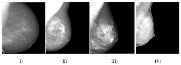
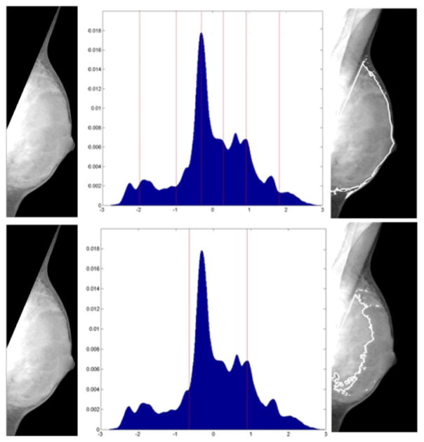
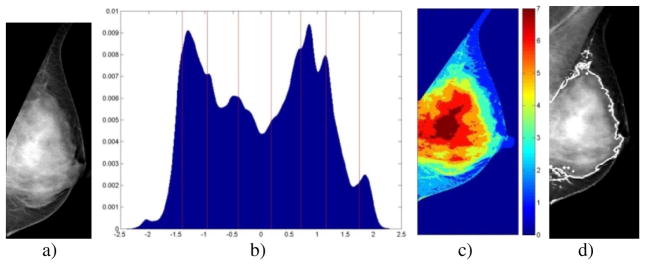
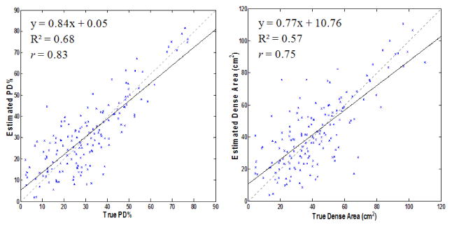
The relative fibroglandular tissue content in the breast, commonly referred to as breast density, has been shown to be the most significant risk factor for breast cancer after age. Currently, the most common approaches to quantify density are based on either semi-automated methods or visual assessment, both of which are highly subjective. This work presents a novel multi-class fuzzy c-means (FCM) algorithm for fully-automated identification and quantification of breast density, optimized for the imaging characteristics of digital mammography. The proposed algorithm involves adaptive FCM clustering based on an optimal number of clusters derived by the tissue properties of the specific mammogram, followed by generation of a final segmentation through cluster agglomeration using linear discriminant analysis. When evaluated on 80 bilateral screening digital mammograms, a strong correlation was observed between algorithm-estimated PD% and radiological ground-truth of r=0.83 (p<0.001) and an average Jaccard spatial similarity coefficient of 0.62. These results show promise for the clinical application of the algorithm in quantifying breast density in a repeatable manner.

摘要

乳房中的相对纤维腺体组织含量,通常称为乳房密度,已被证明是年龄增长后乳腺癌最重要的风险因素。目前,量化密度最常用的方法是基于半自动方法或视觉评估,这两种方法都具有高度主观性。这项工作提出了一种新颖的多类模糊c均值(FCM)算法,用于全自动识别和量化乳房密度,并针对数字乳腺摄影的成像特性进行了优化。所提出的算法包括基于特定乳房X光片的组织特性得出的最佳聚类数进行自适应FCM聚类,然后使用线性判别分析通过聚类合并生成最终分割。在80幅双侧筛查数字乳腺摄影图像上进行评估时,观察到算法估计的PD%与放射学真值之间存在很强的相关性,r = 0.83(p < 0.001),平均杰卡德空间相似系数为0.62。这些结果表明该算法在以可重复的方式量化乳房密度方面具有临床应用前景。

https://cdn.ncbi.nlm.nih.gov/pmc/blobs/a0f5/5510350/5db8f5b2da55/nihms872336f4.jpg
https://cdn.ncbi.nlm.nih.gov/pmc/blobs/a0f5/5510350/fff996ee053a/nihms872336f1.jpg
https://cdn.ncbi.nlm.nih.gov/pmc/blobs/a0f5/5510350/4da7760e8be0/nihms872336f2.jpg
https://cdn.ncbi.nlm.nih.gov/pmc/blobs/a0f5/5510350/476a78f6328a/nihms872336f3.jpg
https://cdn.ncbi.nlm.nih.gov/pmc/blobs/a0f5/5510350/5db8f5b2da55/nihms872336f4.jpg

相似文献

1
Adaptive multi-cluster fuzzy C-means segmentation of breast parenchymal tissue in digital mammography.数字乳腺摄影中乳腺实质组织的自适应多簇模糊C均值分割
Med Image Comput Comput Assist Interv. 2011;14(Pt 3):562-9. doi: 10.1007/978-3-642-23626-6_69.
2
Estimation of breast percent density in raw and processed full field digital mammography images via adaptive fuzzy c-means clustering and support vector machine segmentation.通过自适应模糊C均值聚类和支持向量机分割法估计原始及处理后的全视野数字化乳腺摄影图像中的乳腺密度百分比
Med Phys. 2012 Aug;39(8):4903-17. doi: 10.1118/1.4736530.
3
Automated fibroglandular tissue segmentation and volumetric density estimation in breast MRI using an atlas-aided fuzzy C-means method.基于图谱辅助模糊 C 均值法的乳腺 MRI 中纤维腺体组织自动分割及容积密度估测
Med Phys. 2013 Dec;40(12):122302. doi: 10.1118/1.4829496.
4
An Efficient Segmentation and Classification System in Medical Images Using Intuitionist Possibilistic Fuzzy C-Mean Clustering and Fuzzy SVM Algorithm.基于直觉模糊可能性聚类和模糊 SVM 算法的医学图像高效分割与分类系统。
Sensors (Basel). 2020 Jul 13;20(14):3903. doi: 10.3390/s20143903.
5
Development of a quantitative method for analysis of breast density based on three-dimensional breast MRI.基于三维乳腺磁共振成像的乳腺密度定量分析方法的开发。
Med Phys. 2008 Dec;35(12):5253-62. doi: 10.1118/1.3002306.
6
Computerized calculation of breast density: our experience from Arcadia Medical Imaging Center.乳腺密度的计算机化计算:我们在阿卡迪亚医学影像中心的经验。
Rev Med Chir Soc Med Nat Iasi. 2014 Oct-Dec;118(4):979-85.
7
Geometry-Based Pectoral Muscle Segmentation From MLO Mammogram Views.基于几何方法的MLO位乳房X线照片视图中胸肌分割
IEEE Trans Biomed Eng. 2017 Nov;64(11):2662-2671. doi: 10.1109/TBME.2017.2649481.
8
Suspicious Lesion Segmentation on Brain, Mammograms and Breast MR Images Using New Optimized Spatial Feature Based Super-Pixel Fuzzy C-Means Clustering.基于新型优化空间特征的超像素模糊 C 均值聚类的脑、乳腺 X 线摄影术及乳腺磁共振图像可疑病变分割。
J Digit Imaging. 2019 Apr;32(2):322-335. doi: 10.1007/s10278-018-0149-9.
9
Location of mammograms ROI's and reduction of false-positive.乳房X光检查感兴趣区域的定位及假阳性的减少
Comput Methods Programs Biomed. 2017 May;143:97-111. doi: 10.1016/j.cmpb.2017.02.003. Epub 2017 Feb 24.
10
Radiomics based detection and characterization of suspicious lesions on full field digital mammograms.基于放射组学的全数字化乳腺摄影中可疑病灶的检测与特征描述。
Comput Methods Programs Biomed. 2018 Sep;163:1-20. doi: 10.1016/j.cmpb.2018.05.017. Epub 2018 May 18.

引用本文的文献

1
Complete, Fully Automatic Detection and Classification of Benign and Malignant Breast Tumors Based on CT Images Using Artificial Intelligent and Image Processing.基于人工智能和图像处理技术,利用CT图像对乳腺良恶性肿瘤进行完整、全自动检测与分类
J Clin Med. 2023 Feb 16;12(4):1582. doi: 10.3390/jcm12041582.
2
Breast Cancer Detection using Crow Search Optimization based Intuitionistic Fuzzy Clustering with Neighborhood Attraction.基于具有邻域吸引力的乌鸦搜索优化直觉模糊聚类的乳腺癌检测
Asian Pac J Cancer Prev. 2019 Jan 25;20(1):157-165. doi: 10.31557/APJCP.2019.20.1.157.
3
Automatic and fast segmentation of breast region-of-interest (ROI) and density in MRIs.

本文引用的文献

1
Screening and prevention of breast cancer in primary care.基层医疗中乳腺癌的筛查与预防
Prim Care. 2009 Sep;36(3):533-58. doi: 10.1016/j.pop.2009.04.003.
2
Breast percent density: estimation on digital mammograms and central tomosynthesis projections.乳腺密度百分比:数字乳腺X线摄影及乳腺断层合成中心投照影像的评估
Radiology. 2009 Jul;252(1):40-9. doi: 10.1148/radiol.2521081621. Epub 2009 May 6.
3
Mammographic density estimation: comparison among BI-RADS categories, a semi-automated software and a fully automated one.乳腺X线密度估计:BI-RADS分类、半自动软件和全自动软件之间的比较
磁共振成像中乳腺感兴趣区域(ROI)及密度的自动快速分割
Heliyon. 2018 Dec 17;4(12):e01042. doi: 10.1016/j.heliyon.2018.e01042. eCollection 2018 Dec.
4
Integrating mammographic breast density in glandular dose calculation.将乳腺钼靶密度纳入腺体剂量计算。
Br J Radiol. 2018 May;91(1085):20180032. doi: 10.1259/bjr.20180032. Epub 2018 Feb 13.
5
Effect of Vitamin D Supplementation on Breast Cancer Biomarkers: CALGB 70806 (Alliance) Study Design and Baseline Data.补充维生素D对乳腺癌生物标志物的影响:CALGB 70806(联盟)研究设计与基线数据
Am J Hematol Oncol. 2016 Jul;12(7):4-9.
6
The Impact of Acquisition Dose on Quantitative Breast Density Estimation with Digital Mammography: Results from ACRIN PA 4006.采集剂量对数字化乳腺摄影定量乳腺密度估计的影响:来自美国放射学会影像网络(ACRIN)PA 4006研究的结果
Radiology. 2016 Sep;280(3):693-700. doi: 10.1148/radiol.2016151749. Epub 2016 Mar 22.
7
A Review on Automatic Mammographic Density and Parenchymal Segmentation.乳腺X线摄影密度自动检测与实质分割综述
Int J Breast Cancer. 2015;2015:276217. doi: 10.1155/2015/276217. Epub 2015 Jun 11.
8
AutoDensity: an automated method to measure mammographic breast density that predicts breast cancer risk and screening outcomes.自动密度:一种测量乳腺钼靶密度的自动化方法,可预测乳腺癌风险和筛查结果。
Breast Cancer Res. 2013;15(5):R80. doi: 10.1186/bcr3474.
9
Mammographic parenchymal patterns as an imaging marker of endogenous hormonal exposure: a preliminary study in a high-risk population.乳腺实质类型作为内源性激素暴露的影像学标志物:高危人群的初步研究。
Acad Radiol. 2013 May;20(5):635-46. doi: 10.1016/j.acra.2012.12.016.
10
Estimation of breast percent density in raw and processed full field digital mammography images via adaptive fuzzy c-means clustering and support vector machine segmentation.通过自适应模糊C均值聚类和支持向量机分割法估计原始及处理后的全视野数字化乳腺摄影图像中的乳腺密度百分比
Med Phys. 2012 Aug;39(8):4903-17. doi: 10.1118/1.4736530.
Breast. 2009 Feb;18(1):35-40. doi: 10.1016/j.breast.2008.09.005. Epub 2008 Nov 17.
4
Mammographic density. Measurement of mammographic density.乳腺X线密度。乳腺X线密度的测量。
Breast Cancer Res. 2008;10(3):209. doi: 10.1186/bcr2102. Epub 2008 Jun 19.
5
A novel breast tissue density classification methodology.一种新型的乳腺组织密度分类方法。
IEEE Trans Inf Technol Biomed. 2008 Jan;12(1):55-65. doi: 10.1109/TITB.2007.903514.
6
Basic physics and doubts about relationship between mammographically determined tissue density and breast cancer risk.基础物理学以及关于乳腺钼靶检查确定的组织密度与乳腺癌风险之间关系的疑问。
Radiology. 2008 Feb;246(2):348-53. doi: 10.1148/radiol.2461070309.
7
Computing mammographic density from a multiple regression model constructed with image-acquisition parameters from a full-field digital mammographic unit.根据由全视野数字乳腺摄影设备的图像采集参数构建的多元回归模型计算乳腺X线摄影密度。
Phys Med Biol. 2007 Aug 21;52(16):4905-21. doi: 10.1088/0031-9155/52/16/013. Epub 2007 Jul 30.
8
Accuracy of assigned BI-RADS breast density category definitions.指定的乳腺影像报告和数据系统(BI-RADS)乳腺密度分类定义的准确性。
Acad Radiol. 2006 Sep;13(9):1143-9. doi: 10.1016/j.acra.2006.06.005.
9
Mammographic density measured with quantitative computer-aided method: comparison with radiologists' estimates and BI-RADS categories.采用定量计算机辅助方法测量的乳腺密度:与放射科医生的评估及BI-RADS分类的比较。
Radiology. 2006 Sep;240(3):656-65. doi: 10.1148/radiol.2402041947. Epub 2006 Jul 20.
10
Automated classification of parenchymal patterns in mammograms.乳腺钼靶片中实质模式的自动分类。
Phys Med Biol. 1998 Feb;43(2):365-78. doi: 10.1088/0031-9155/43/2/011.