Shrestha Bhushan, Han Sang-Kuk, Yoon Kwon-Sang, Sung Jae-Mo
Entomopathogenic Fungal Culture Collection, Department of Applied Biology, Kangwon National University, Chuncheon 200-701, Korea.
Mycobiology. 2005 Jun;33(2):69-76. doi: 10.4489/MYCO.2005.33.2.069. Epub 2005 Jun 30.
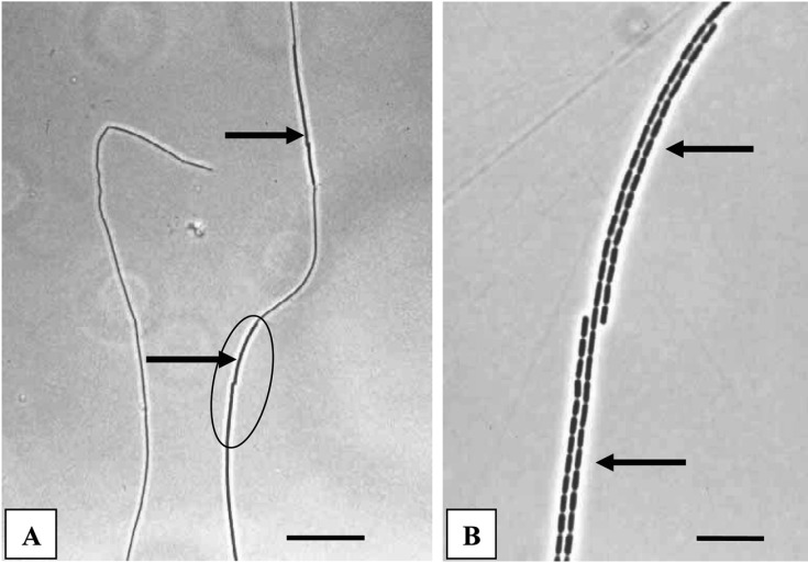
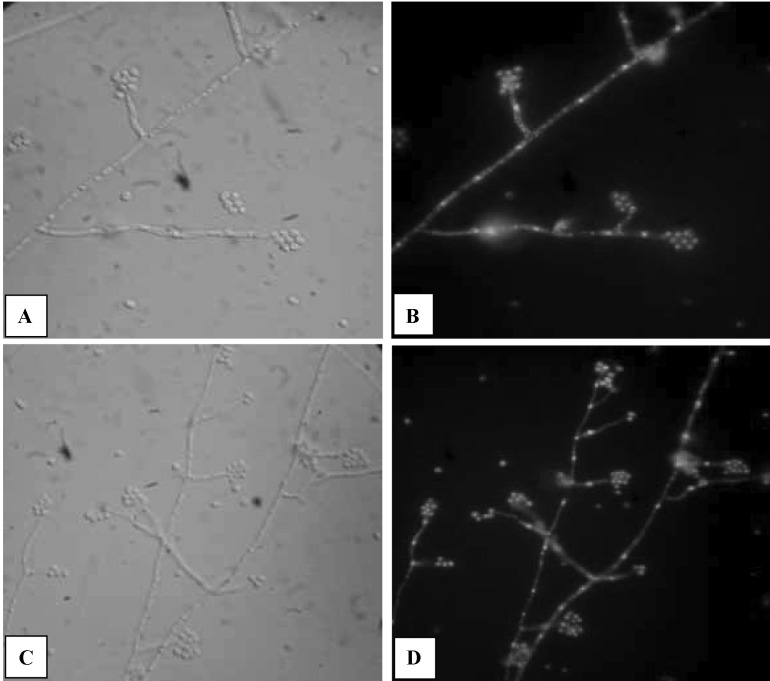
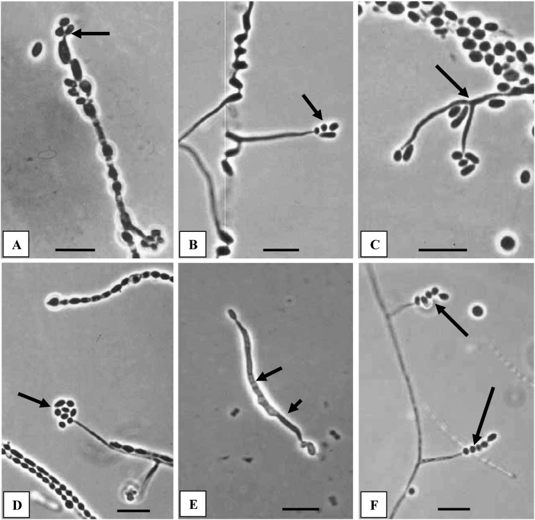
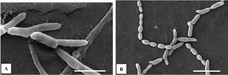
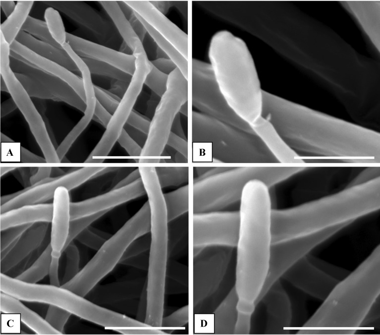
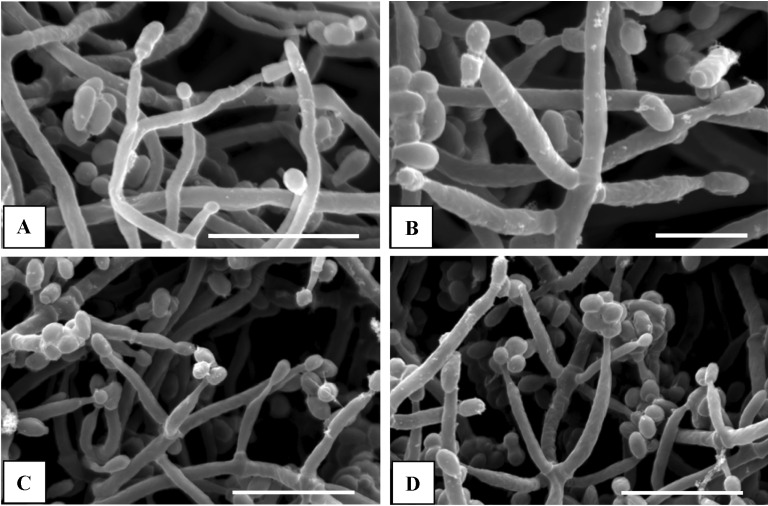
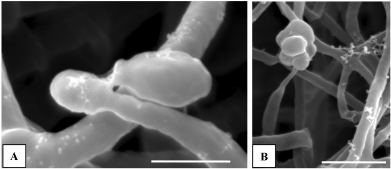
Conidial development of Cordyceps militaris was observed from germinating ascospores and vegetative hyphae through light and scanning electron microscopy (SEM). Ascospores were discharged from fresh specimens of C. militaris in sterile water as well as Sabouraud Dextrose agar plus Yeast Extract (SDAY) plates. We observed ascospore germination and conidial formation periodically. Under submerged condition in sterile water, most part-spores germinated unidirectionally and conidia were developed directly from the tips of germinating hyphae of part-spores within 36 h after ascospore discharge, showing microcyclic conidiation. First-formed conidia were cylindrical or clavate followed by globose and ellipsoidal ones. Germination of ascospores and conidial development were observed on SDAY agar by SEM. Slimy heads of conidia on variously arranged phialides, from solitary to whorl, developed 5 days after ascospore discharge. Besides, two distinct types of conidia, elongated pyriform or cylindrical and globose, were observed in the same slimy heads by SEM. Conidia were shown to be uninucleate with 4,6-diamidino-2-phenylindole staining. Conidiogenous cells were more slender than vegetative hyphae, having attenuated tips. Microcyclic conidiation, undifferentiated conidiogenous hyphae (phialides), polymorphic conidia and solitary, opposite to whorled type of phialidic arrangement are reported here as the characteristic features of asexual stage of C. militaris, which can be distinguished from other Cordyceps species.
通过光学显微镜和扫描电子显微镜(SEM)观察了北虫草的分生孢子发育过程,观察对象包括萌发的子囊孢子和营养菌丝。将新鲜的北虫草标本中的子囊孢子接种于无菌水以及含酵母提取物的沙氏葡萄糖琼脂(SDAY)平板上。我们定期观察子囊孢子的萌发和分生孢子的形成。在无菌水的浸没条件下,大部分分孢子单向萌发,分生孢子在子囊孢子释放后36小时内直接从分孢子萌发菌丝的顶端发育形成,呈现出微循环产孢。最初形成的分生孢子为圆柱形或棒状,随后是球形和椭圆形。通过扫描电子显微镜观察了SDAY琼脂上子囊孢子的萌发和分生孢子的发育情况。在子囊孢子释放5天后,在各种排列方式的瓶梗上(从单个到轮生)形成了分生孢子的黏液头。此外,通过扫描电子显微镜在同一黏液头中观察到了两种不同类型的分生孢子,即细长梨形或圆柱形以及球形。经4,6-二脒基-2-苯基吲哚染色显示分生孢子为单核。产孢细胞比营养菌丝更细长,顶端逐渐变细。本文报道了微循环产孢、未分化的产孢菌丝(瓶梗)、多态性分生孢子以及瓶梗排列方式为单个、对生到轮生,这些是北虫草无性阶段的特征,可将其与其他虫草种类区分开来。