Song Yafeng, Stål Per S, Yu Jiguo, Forsgren Sture
Section for Anatomy, Department of Integrative Medical Biology, Umeå University, 901 87 Umeå, Sweden.
ISRN Inflamm. 2013 Jan 29;2013:907821. doi: 10.1155/2013/907821. eCollection 2013.
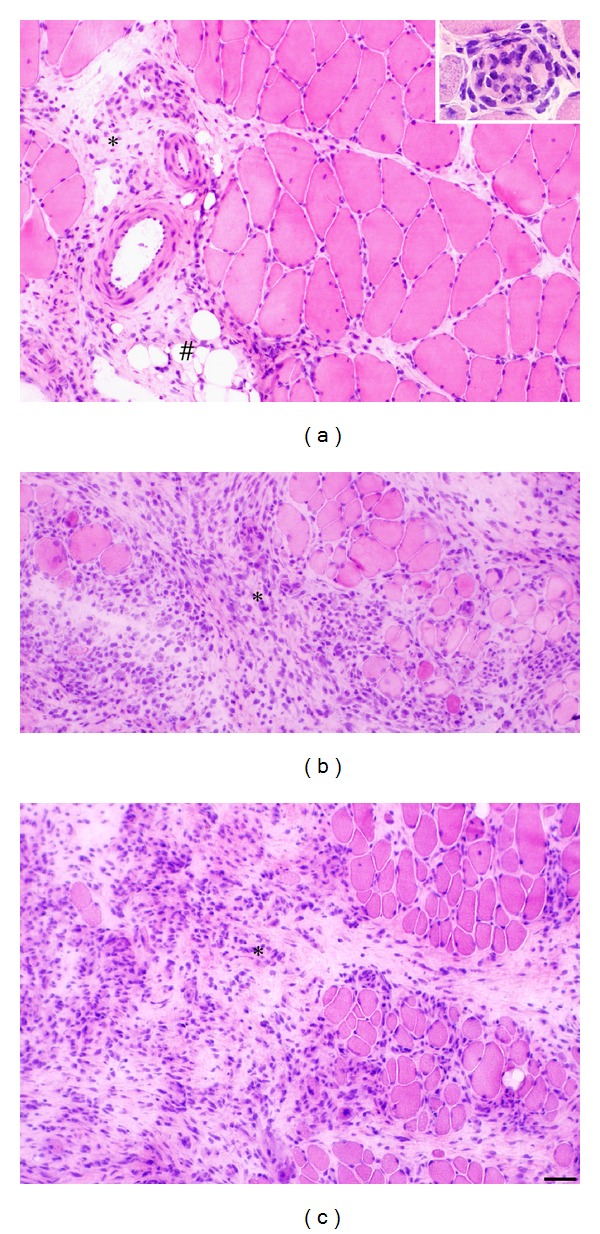
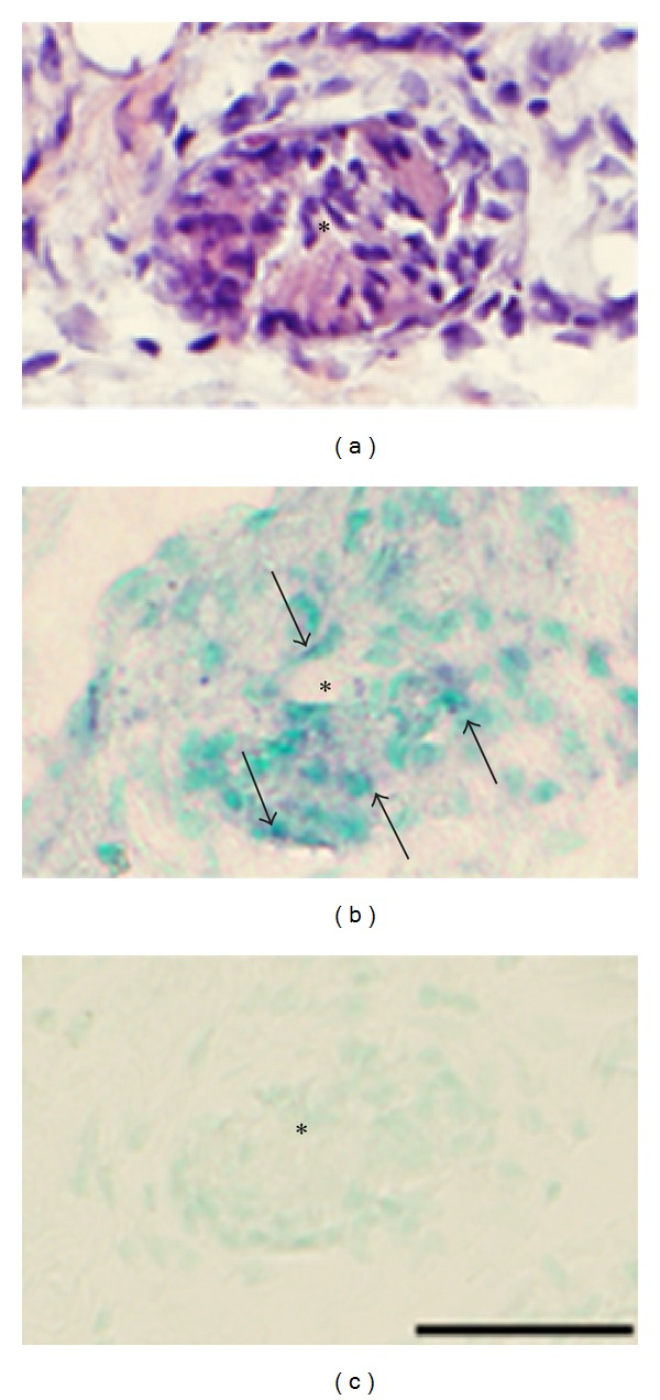
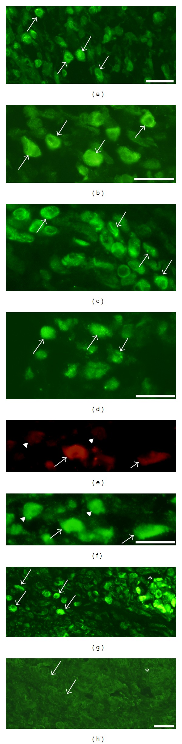
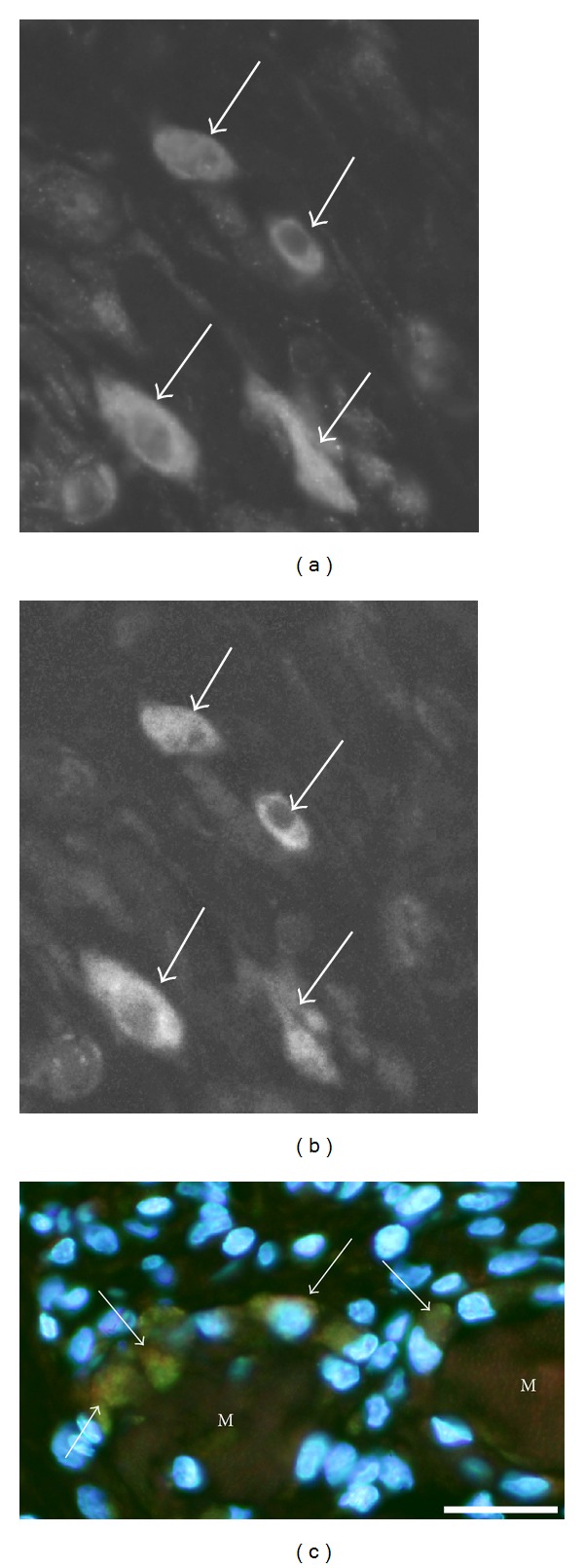
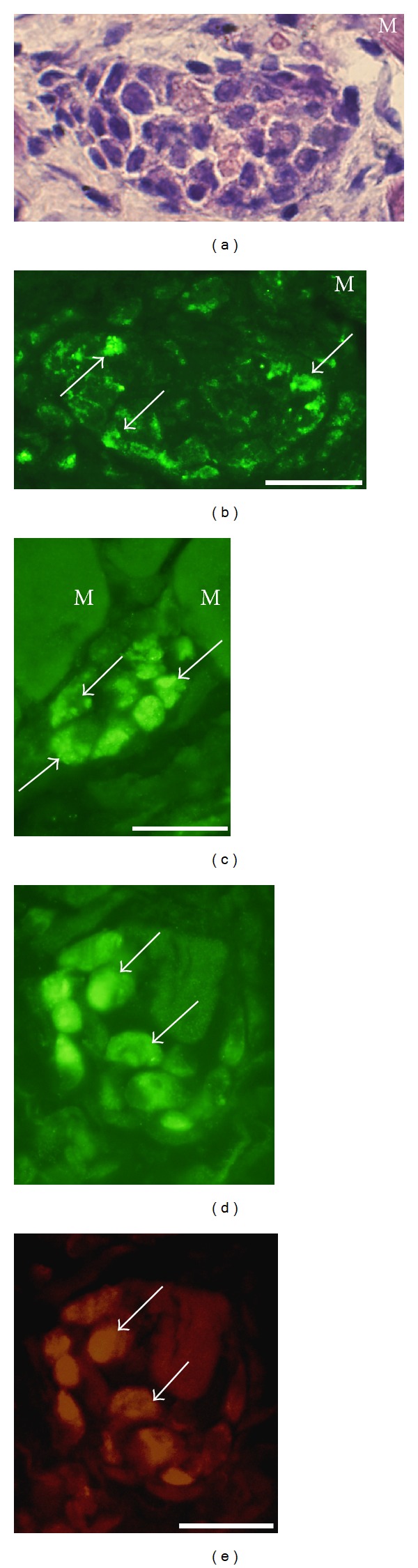
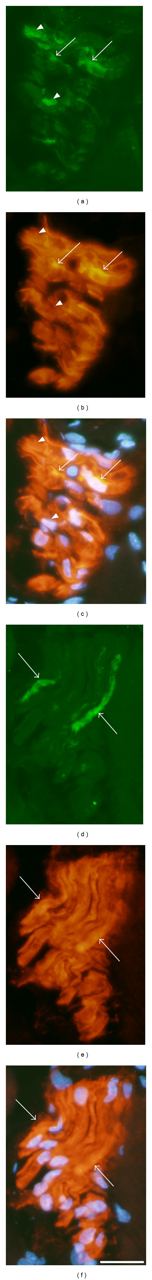
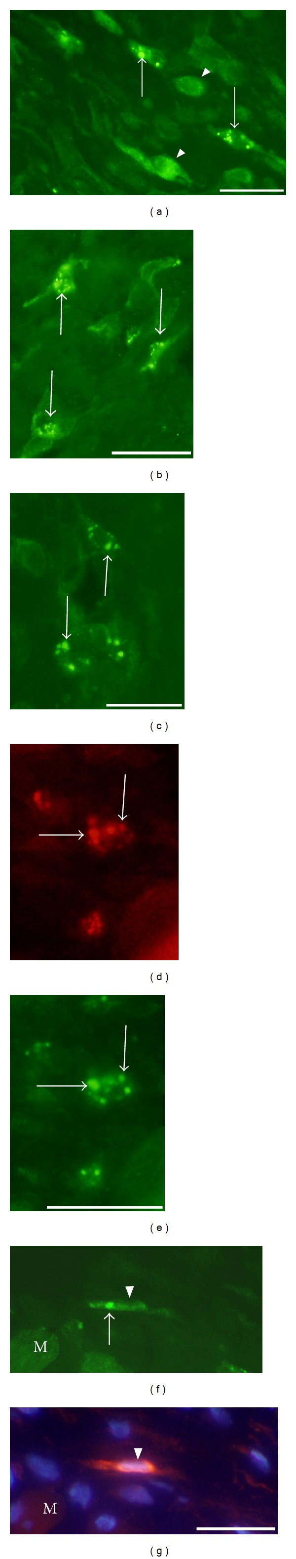
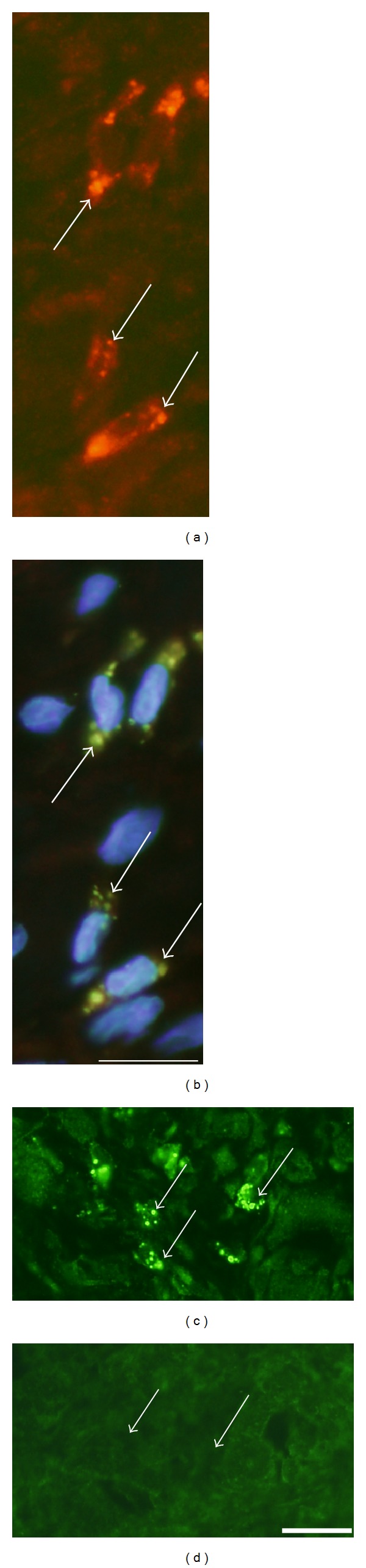
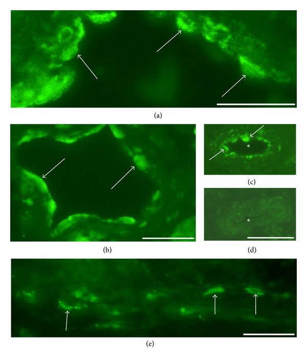
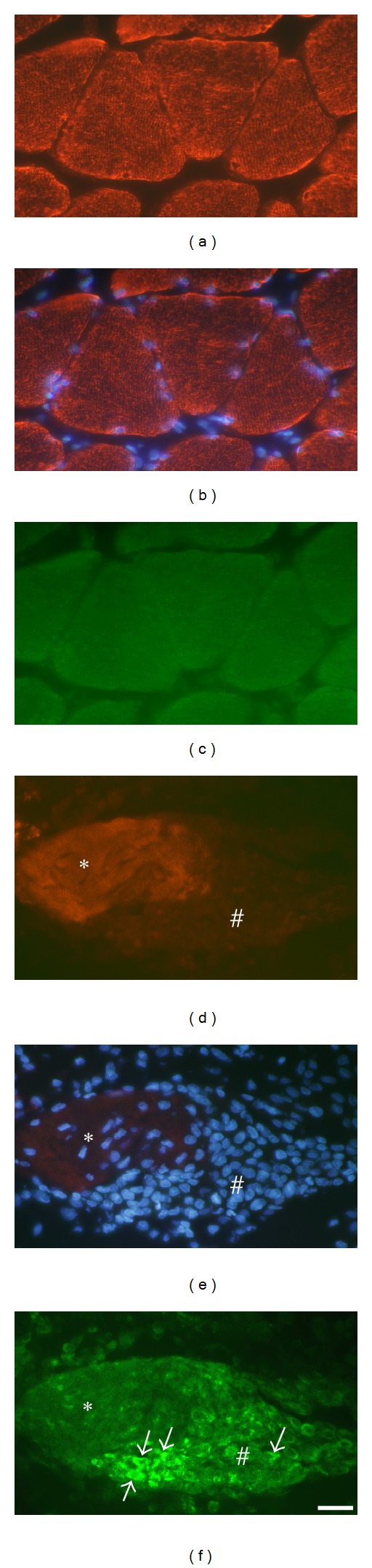
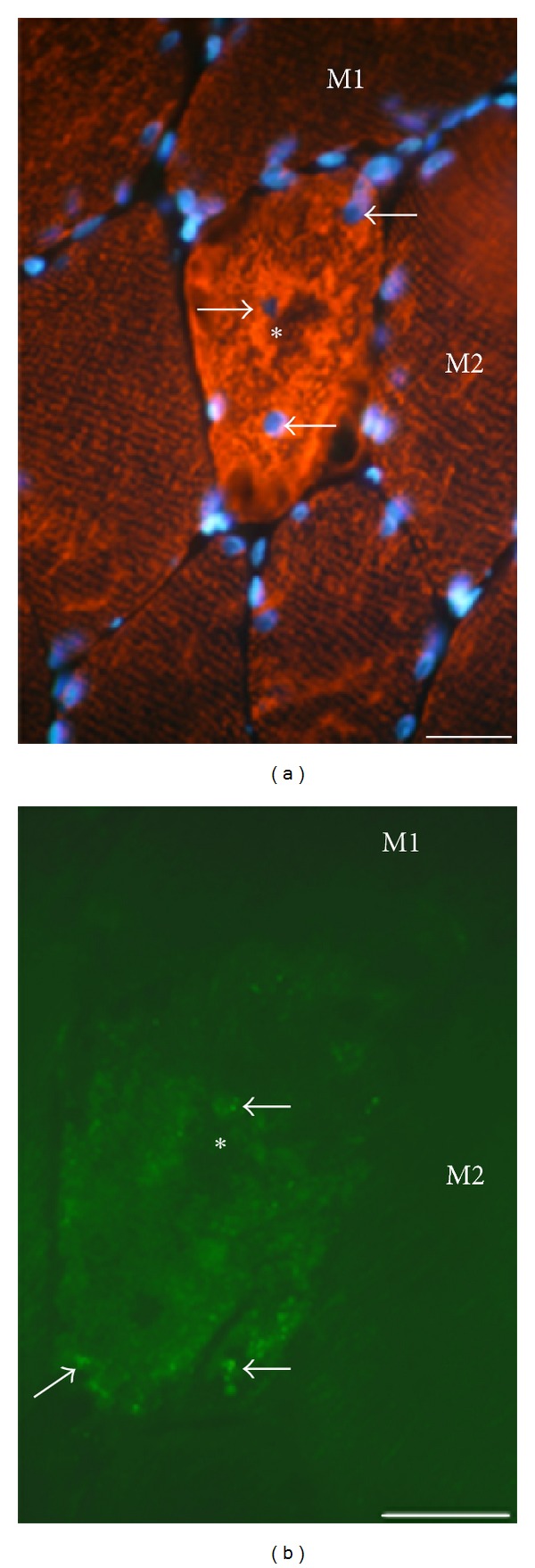
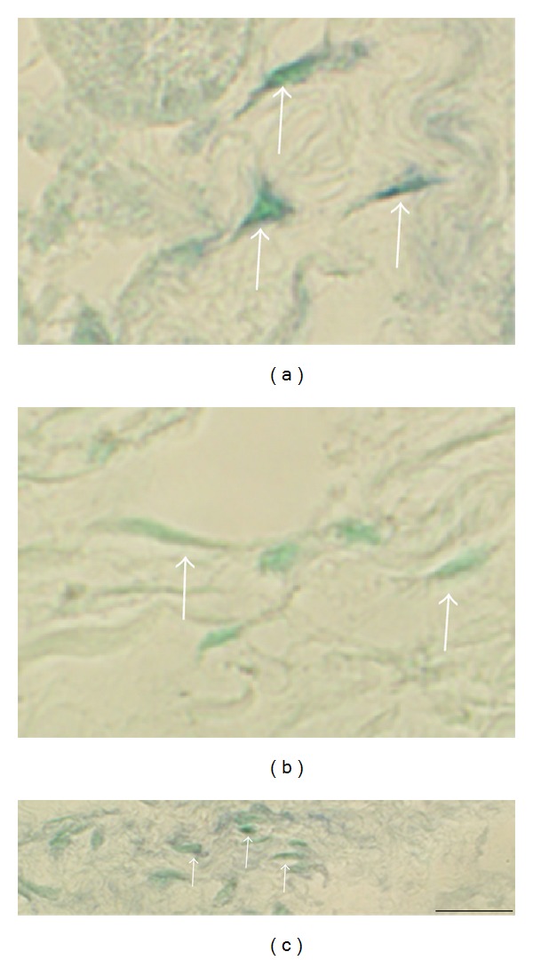
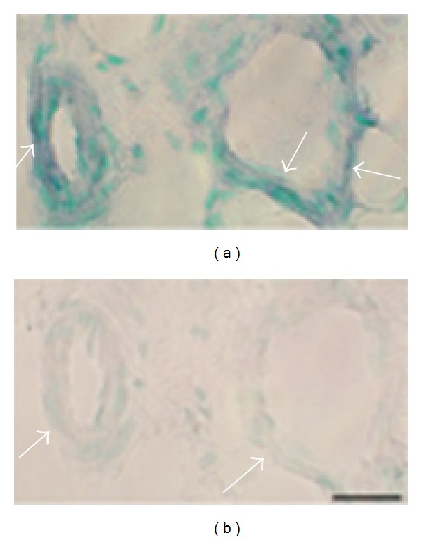
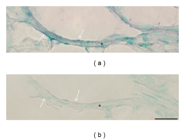
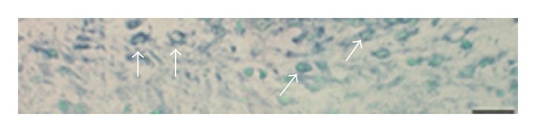
Muscle injury and inflammation (myositis) in a rabbit model of an unilateral muscle overuse were examined. It is unknown if the tachykinin system has a functional role in this situation. In this study, therefore, the neurokinin-1 receptor (NK-1R) expression patterns were evaluated. White blood cells, nerve fascicles, fine nerve fibers, and blood vessel walls in myositis areas showed NK-1R immunoreaction. NK-1R mRNA reactions were observable for white blood cells and blood vessel walls of these areas. NK-1R immunoreaction and NK-1R mRNA reactions were also seen for muscle fibers showing degenerative and regenerative features. There were almost no NK-1R immunoreactions in normal muscle tissue. Interestingly, marked NK-1R expressions were seen for myositis areas of both the experimental side and the contralateral nonexperimental side. EIA analyses showed that the concentration of substance P in the muscle tissue was clearly increased bilaterally at the experimental end stage, as compared to the situation for normal muscle tissue. These observations show that the tachykinin system is very much involved in the processes that occur in muscle injury/myositis. The effects can be related to proinflammatory effects and/or tissue repair. The fact that there are also marked NK-1R expressions contralaterally indicate that the tachykinin system has crossover effects.
在单侧肌肉过度使用的兔模型中,对肌肉损伤和炎症(肌炎)进行了检查。目前尚不清楚速激肽系统在这种情况下是否具有功能性作用。因此,在本研究中,对神经激肽-1受体(NK-1R)的表达模式进行了评估。肌炎区域的白细胞、神经束、细神经纤维和血管壁显示出NK-1R免疫反应。在这些区域的白细胞和血管壁中可观察到NK-1R mRNA反应。在显示退行性和再生特征的肌纤维中也可见到NK-1R免疫反应和NK-1R mRNA反应。正常肌肉组织中几乎没有NK-1R免疫反应。有趣的是,在实验侧和对侧非实验侧的肌炎区域均可见明显的NK-1R表达。酶免疫分析表明,与正常肌肉组织相比,在实验末期,肌肉组织中P物质的浓度在双侧均明显升高。这些观察结果表明,速激肽系统在很大程度上参与了肌肉损伤/肌炎中发生的过程。其作用可能与促炎作用和/或组织修复有关。对侧也有明显的NK-1R表达这一事实表明速激肽系统具有交叉效应。