Experimental Biophysics and Applied Nanoscience, Faculty of Physics and Bielefeld Institute for Biophysics and Nanoscience (BINAS), Bielefeld University, Universitätsstrasse 25, D-33615 Bielefeld, Germany.
Beilstein J Nanotechnol. 2013 Sep 11;4:510-6. doi: 10.3762/bjnano.4.60. eCollection 2013.
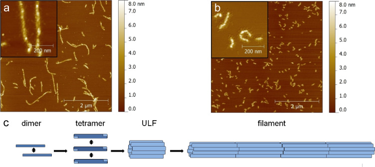
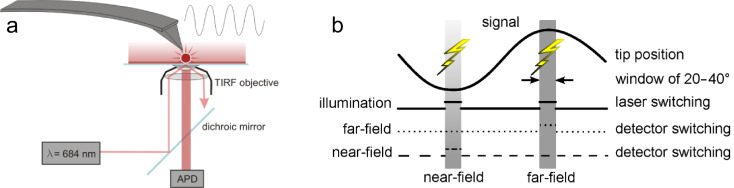
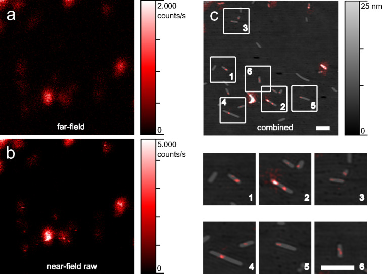
Both fluorescence imaging and atomic force microscopy (AFM) are highly versatile and extensively used in applications ranging from nanotechnology to life sciences. In fluorescence microscopy luminescent dyes serve as position markers. Moreover, they can be used as active reporters of their local vicinity. The dipolar coupling of the tip with the incident light and the fluorophore give rise to a local field and fluorescence enhancement. AFM topographic imaging allows for resolutions down to the atomic scale. It can be operated in vacuum, under ambient conditions and in liquids. This makes it ideal for the investigation of a wide range of different samples. Furthermore an illuminated AFM cantilever tip apex exposes strongly confined non-propagating electromagnetic fields that can serve as a coupling agent for single dye molecules. Thus, combining both techniques by means of apertureless scanning near-field optical microscopy (aSNOM) enables concurrent high resolution topography and fluorescence imaging. Commonly, among the various (apertureless) SNOM approaches metallic or metallized probes are used. Here, we report on our custom-built aSNOM setup, which uses commercially available monolithic silicon AFM cantilevers. The field enhancement confined to the tip apex facilitates an optical resolution down to 20 nm. Furthermore, the use of standard mass-produced AFM cantilevers spares elaborate probe production or modification processes. We investigated tobacco mosaic viruses and the intermediate filament protein desmin. Both are mixed complexes of building blocks, which are fluorescently labeled to a low degree. The simultaneous recording of topography and fluorescence data allows for the exact localization of distinct building blocks within the superordinate structures.
荧光成像和原子力显微镜(AFM)都是非常通用的,广泛应用于从纳米技术到生命科学的各个领域。在荧光显微镜中,荧光染料用作位置标记物。此外,它们可以用作其局部环境的主动示踪剂。尖端与入射光和荧光团的偶极耦合会产生局部场和荧光增强。AFM 形貌成像可以达到原子级分辨率。它可以在真空中、环境条件下和液体中操作。这使其成为研究各种不同样品的理想选择。此外,照明的 AFM 悬臂尖端会暴露强烈受限的非传播电磁场,这些电磁场可用作单染料分子的耦合剂。因此,通过无孔扫描近场光学显微镜(aSNOM)将这两种技术结合起来,可以实现高分辨率形貌和荧光同时成像。通常,在各种(无孔)SNOM 方法中,使用金属或金属化的探针。在这里,我们报告了我们定制的 aSNOM 装置,它使用市售的整体式硅 AFM 悬臂。尖端的场增强有助于将光学分辨率降低到 20nm。此外,使用标准批量生产的 AFM 悬臂可以省去复杂的探针制造或修改过程。我们研究了烟草花叶病毒和中间丝蛋白结蛋白。这两种都是混合构建块的复合物,它们的荧光标记程度较低。同时记录形貌和荧光数据可以精确定位超结构内的不同构建块。