Positron Emission Tomography Centre, IRMET S.p.A., Euromedic Inc., 10136 Turin, Italy ; Co-ordinator of PET Pediatric AIMN InterGroup, 10136 Turin, Italy ; Associate researcher of Institute of Cognitive Sciences and Technologies, National Research Council, 00185 Rome, Italy.
Biomed Res Int. 2013;2013:709037. doi: 10.1155/2013/709037. Epub 2013 Aug 26.
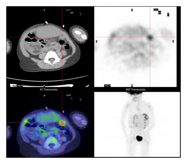
In this study we retrospectively evaluated if ¹⁸F-FDG-PET/CT provided incremental diagnostic information over CI in a group of hepatoblastoma patients performing restaging.
Nine patients (mean age: 5.9 years; range: 3.1-12 years) surgically treated for hepatoblastoma were followed up by clinical examination, serum α-FP monitoring, and US. CI (CT or MRI) and PET/CT were performed in case of suspicion of relapse. Fine-needle aspiration biopsies (FNAB) were carried out for final confirmation if the results of CI, PET/CT, and/or α-FP levels were suggestive of relapse. PET/CT and CI findings were analyzed for comparison purposes, using FNAB as reference standard.
α-FP level was suggestive of disease recurrence in 8/9 patients. Biopsy was performed in 8/9 cases. CI and PET/CT resulted to be concordant in 5/9 patients (CI identified recurrence of disease, but ¹⁸F-FDG-PET/CT provided a better definition of disease extent); in 4/9 cases, CI diagnostic information resulted in negative findings, whereas PET/CT correctly detected recurrence of disease. ¹⁸F-FDG-PET/CT showed an agreement of 100% (8/8) with FNAB results.
¹⁸F-FDG-PET/CT scan seems to better assess HB patients with respect to CI and may provide incremental diagnostic value in the restaging of this group of patients.
在本研究中,我们回顾性评估了¹⁸F-FDG-PET/CT 在一组接受再分期的肝母细胞瘤患者中是否比 CI 提供了额外的诊断信息。
9 例(平均年龄:5.9 岁;范围:3.1-12 岁)接受肝母细胞瘤手术治疗的患者接受临床检查、血清α-FP 监测和 US 随访。如果怀疑复发,将进行 CI(CT 或 MRI)和 PET/CT。如果 CI、PET/CT 和/或α-FP 水平提示复发,将进行细针抽吸活检(FNAB)以最终确认。
8/9 例患者的α-FP 水平提示疾病复发。8/9 例患者均进行了活检。为了比较目的,对 PET/CT 和 CI 结果进行了分析,以 FNAB 为参考标准。在 5/9 例患者中,CI 和 PET/CT 的结果是一致的(CI 发现疾病复发,但¹⁸F-FDG-PET/CT 提供了更好的疾病范围定义);在 4/9 例患者中,CI 的诊断信息结果为阴性,但 PET/CT 正确地检测到疾病复发。¹⁸F-FDG-PET/CT 与 FNAB 结果的一致性为 100%(8/8)。
¹⁸F-FDG-PET/CT 扫描似乎比 CI 更好地评估 HB 患者,并且可能在该组患者的再分期中提供额外的诊断价值。