Center for Medical Imaging and Physiology, Skåne University Hospital, Lund, Lund University, 221 85, Lund, Sweden,
Insights Imaging. 2013 Dec;4(6):787-98. doi: 10.1007/s13244-013-0288-y. Epub 2013 Sep 25.
The first radiographic scoring system for pulmonary cystic fibrosis was presented in 1958. Since then a multitude of scoring systems for radiography and computed tomography (CT) have been presented, recently also for tomosynthesis and magnetic resonance imaging (MRI). The aim of the current review was to analyse and compare the plethora of scoring systems for cystic fibrosis, especially regarding which scoring components are considered most important.
Four scoring systems for chest radiography, one for tomosynthesis, eight for CT and one for MRI were compared regarding components evaluated and their terminology; the areas scored; scoring levels; the weighting of each component in percentage of the total score; and the calculations for the final score.
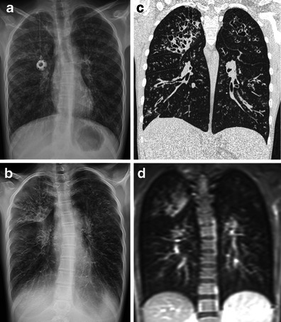
In most radiological scoring systems the lungs are evaluated for increased volume, bronchial wall thickening, bronchiectasis, mucus plugging, atelectasis and consolidation. In addition, for instance abscesses, bullae, septal thickening, mosaic perfusion, ground glass opacities and air trapping are evaluated in some CT scoring systems. Pleural affection and perfusion defects are scored on MRI.
Bronchiectasis alone, or in combination with mucus plugging, is given the highest weighting in most scoring systems and is thus commonly considered to be the most significant finding when evaluating cystic fibrosis lung disease.
• Scoring of examinations is used for comparison of outcome in studies. • Scoring of examinations can also be used for monitoring disease progression. • Cystic fibrosis can be scored on radiography, tomosynthesis, CT or MRI. • The typical imaging findings of cystic fibrosis depend on the imaging modality used. • Bronchiectasis is commonly considered the most significant finding when scoring cystic fibrosis.
1958 年提出了首个用于囊性纤维化肺部的放射学评分系统。此后,已经提出了许多用于放射摄影和计算机断层扫描(CT)的评分系统,最近还提出了断层合成和磁共振成像(MRI)的评分系统。本综述的目的是分析和比较大量的囊性纤维化评分系统,特别是评估哪些评分成分最重要。
比较了四种胸部 X 线摄影评分系统、一种断层合成评分系统、八种 CT 评分系统和一种 MRI 评分系统,评估内容及其术语;评分区域;评分级别;每个成分在总分中的权重(百分比);以及最终评分的计算。
在大多数放射学评分系统中,肺部评估包括容积增加、支气管壁增厚、支气管扩张、黏液栓、肺不张和实变。此外,在一些 CT 评分系统中还评估了脓肿、大疱、间隔增厚、马赛克灌注、磨玻璃混浊和空气潴留等。磁共振成像可评估胸膜受累和灌注缺陷。
在大多数评分系统中,支气管扩张单独或与黏液栓一起,权重最高,因此在评估囊性纤维化肺部疾病时通常被认为是最重要的发现。
• 评分可用于研究结果的比较。
• 评分也可用于监测疾病进展。
• 可对放射摄影、断层合成、CT 或 MRI 进行囊性纤维化评分。
• 囊性纤维化的典型影像学表现取决于所使用的成像方式。
• 支气管扩张通常被认为是评分囊性纤维化时最重要的发现。