Radiology and Nuclear Medicine Dept, Erasmus MC, Rotterdam, The Netherlands
Pediatric Pulmonology and Allergology Dept, Erasmus MC, Sophia Children's Hospital, Rotterdam, The Netherlands.
Eur Respir Rev. 2022 Mar 23;31(163). doi: 10.1183/16000617.0173-2021. Print 2022 Mar 31.
Imaging represents an important noninvasive means to assess cystic fibrosis (CF) lung disease, which remains the main cause of morbidity and mortality in CF patients. While the development of new imaging techniques has revolutionised clinical practice, advances have posed diagnostic and monitoring challenges. The authors aim to summarise these challenges and make evidence-based recommendations regarding imaging assessment for both clinicians and radiologists.
A committee of 21 experts in CF from the 10 largest specialist centres in Italy was convened, including a radiologist and a pulmonologist from each centre, with the overall aim of developing clear and actionable recommendations for lung imaging in CF. An threshold of at least 80% of the votes was required for acceptance of each statement of recommendation.
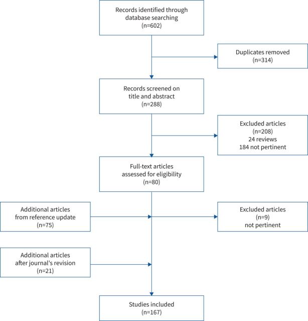
After a systematic review of the relevant literature, the committee convened to evaluate 167 articles. Following five RAND conferences, consensus statements were developed by an executive subcommittee. The entire consensus committee voted and approved 28 main statements.
There is a need for international guidelines regarding the appropriate timing and selection of imaging modality for patients with CF lung disease; timing and selection depends upon the clinical scenario, the patient's age, lung function and type of treatment. Despite its ubiquity, the use of the chest radiograph remains controversial. Both computed tomography and magnetic resonance imaging should be routinely used to monitor CF lung disease. Future studies should focus on imaging protocol harmonisation both for computed tomography and for magnetic resonance imaging. The introduction of artificial intelligence imaging analysis may further revolutionise clinical practice by providing fast and reliable quantitative outcomes to assess disease status. To date, there is no evidence supporting the use of lung ultrasound to monitor CF lung disease.
影像学是评估囊性纤维化(CF)肺部疾病的重要无创手段,该病仍然是 CF 患者发病和死亡的主要原因。虽然新的影像学技术的发展彻底改变了临床实践,但这些进步也带来了诊断和监测方面的挑战。作者旨在总结这些挑战,并就影像学评估为临床医生和放射科医生提供循证建议。
召集了来自意大利最大的 10 个专业中心的 21 名 CF 专家组成一个委员会,每个中心都有一名放射科医生和一名肺病专家,目的是为 CF 肺部成像制定明确且可行的建议。每项推荐声明至少需要 80%的票数才能被接受。
在对相关文献进行系统回顾后,委员会召开会议评估了 167 篇文章。经过五次 RAND 会议,执行小组委员会制定了共识声明。全体共识委员会对 28 项主要声明进行了投票和批准。
需要制定关于 CF 肺部疾病患者的影像学检查的适当时机和选择的国际指南;时机和选择取决于临床情况、患者的年龄、肺功能和治疗类型。尽管胸部 X 线摄影的应用非常广泛,但它的使用仍然存在争议。计算机断层扫描和磁共振成像都应常规用于监测 CF 肺部疾病。未来的研究应侧重于计算机断层扫描和磁共振成像的成像协议的协调。人工智能成像分析的引入可能通过提供快速可靠的定量结果来评估疾病状态,从而进一步彻底改变临床实践。迄今为止,没有证据支持使用肺部超声来监测 CF 肺部疾病。