• 文献检索
  • 文档翻译
  • 深度研究
  • 学术资讯
  • Suppr Zotero 插件Zotero 插件
  • 邀请有礼
  • 套餐&价格
  • 历史记录
应用&插件
Suppr Zotero 插件Zotero 插件浏览器插件Mac 客户端Windows 客户端微信小程序
定价
高级版会员购买积分包购买API积分包
服务
文献检索文档翻译深度研究API 文档MCP 服务
关于我们
关于 Suppr公司介绍联系我们用户协议隐私条款
关注我们

Suppr 超能文献

核心技术专利:CN118964589B侵权必究
粤ICP备2023148730 号-1Suppr @ 2026

文献检索

告别复杂PubMed语法,用中文像聊天一样搜索,搜遍4000万医学文献。AI智能推荐,让科研检索更轻松。

立即免费搜索

文件翻译

保留排版,准确专业,支持PDF/Word/PPT等文件格式,支持 12+语言互译。

免费翻译文档

深度研究

AI帮你快速写综述,25分钟生成高质量综述,智能提取关键信息,辅助科研写作。

立即免费体验

使用漫射光学光谱成像(DOSI)评估乳腺致密组织中的肿瘤对比度。

Assessing tumor contrast in radiographically dense breast tissue using Diffuse Optical Spectroscopic Imaging (DOSI).

作者信息

Leproux Anaïs, Durkin Amanda, Compton Montana, Cerussi Albert E, Gratton Enrico, Tromberg Bruce J

出版信息

Breast Cancer Res. 2013;15(5):R89. doi: 10.1186/bcr3485.

DOI:10.1186/bcr3485
PMID:24066941
原文链接:https://pmc.ncbi.nlm.nih.gov/articles/PMC3979060/
Abstract

INTRODUCTION

Radiographic density adversely affects the performance of X-ray mammography and can be particularly problematic in younger and high-risk women. Because of this limitation, there is significant ongoing effort to develop alternative cancer screening and detection strategies for this population. This pilot study evaluates the potential of Diffuse Optical Spectroscopic Imaging (DOSI) to image known tumors in dense breast tissue.

METHODS

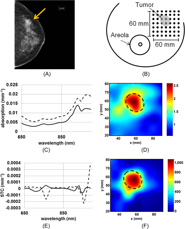
We performed a retrospective analysis on 24 radiographically dense breast cancer subjects measured with DOSI over a four-year period (Breast Imaging Reporting and Data System - BI-RADS, category 3 and 4, average age = 39 ± 7.6, average maximum size 31 ± 1 7 mm). Two previously-described DOSI contrast functions, the tissue optical index (TOI) and the specific tumor component (STC), which are based upon the concentrations and spectral signatures of hemoglobin, water and lipids, respectively, were used to form 2D optical images of breast tumors.

RESULTS

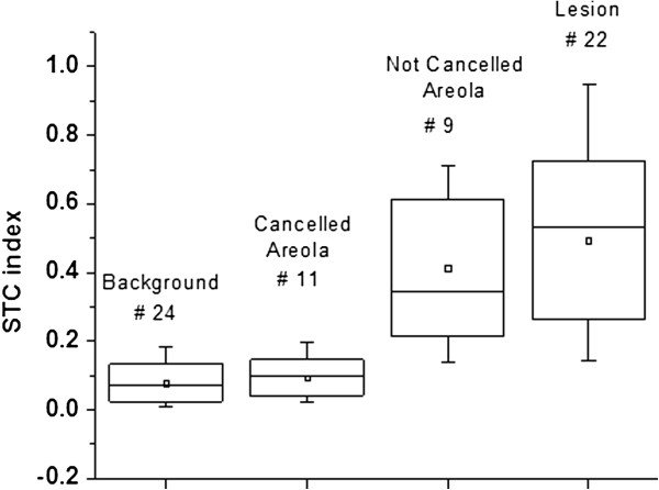
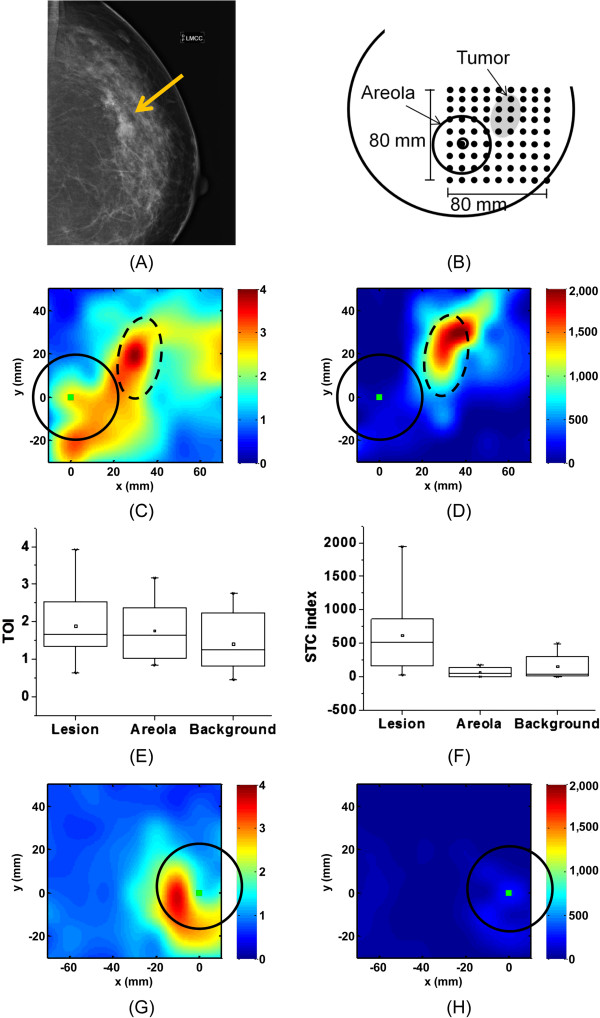
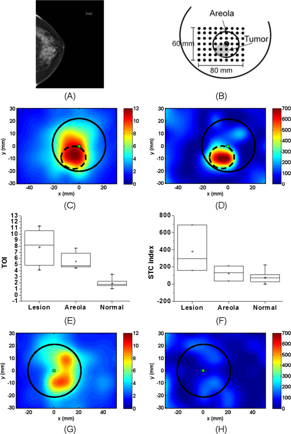
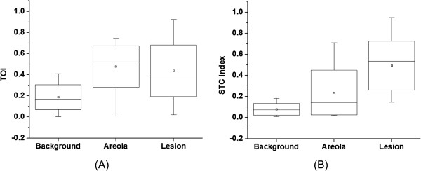
Using TOI and STC, 21 out of 24 breast tumors were found to be statistically different from the surrounding highly vascularized dense tissue and to be distinguishable from the areolar region. For these patients, the tumor to normal contrast was 2.6 ± 1.2 (range 1.3 to 5.5) and 10.0 ± 7.5 (range 3.3 to 26.4) for TOI and STC, respectively. STC images were particularly useful in eliminating metabolic background from the retroareolar region which led to identification of two out of four retroareolar tumors.

CONCLUSIONS

Using both the abundance and the disposition of the tissue chromophores recovered from the DOSI measurements, we were able to observe tumor contrast relative to dense breast tissue. These preliminary results suggest that DOSI spectral characterization strategies may provide new information content that could help imaging breast tumors in radiographically dense tissue and in particular in the areolar complex.

摘要

引言

X线摄影密度会对乳腺X线摄影的性能产生不利影响,在年轻女性和高危女性中问题可能尤为突出。鉴于这一局限性,目前正在大力研发针对该人群的替代癌症筛查和检测策略。本初步研究评估了漫射光学光谱成像(DOSI)对致密乳腺组织中已知肿瘤进行成像的潜力。

方法

我们对24名乳腺X线摄影显示致密的乳腺癌患者进行了回顾性分析,这些患者在四年期间接受了DOSI测量(乳腺影像报告和数据系统-BI-RADS,3类和4类,平均年龄=39±7.6岁,平均最大尺寸31±17mm)。使用两个先前描述的DOSI对比函数,即组织光学指数(TOI)和特定肿瘤成分(STC),分别基于血红蛋白、水和脂质的浓度及光谱特征,来形成乳腺肿瘤的二维光学图像。

结果

使用TOI和STC,24个乳腺肿瘤中有21个在统计学上与周围高度血管化的致密组织不同,并且可与乳晕区域区分开来。对于这些患者,TOI和STC的肿瘤与正常组织的对比度分别为2.6±1.2(范围1.3至5.5)和10.0±7.5(范围3.3至26.4)。STC图像在消除乳晕后区域的代谢背景方面特别有用,这使得四个乳晕后肿瘤中有两个得以识别。

结论

利用从DOSI测量中获得的组织发色团的丰度和分布,我们能够观察到相对于致密乳腺组织的肿瘤对比度。这些初步结果表明,DOSI光谱表征策略可能提供新的信息内容,有助于对致密乳腺组织尤其是乳晕复合体中的乳腺肿瘤进行成像。

https://cdn.ncbi.nlm.nih.gov/pmc/blobs/1ab4/3979060/176ebda239f0/bcr3485-5.jpg
https://cdn.ncbi.nlm.nih.gov/pmc/blobs/1ab4/3979060/bba685853fe9/bcr3485-1.jpg
https://cdn.ncbi.nlm.nih.gov/pmc/blobs/1ab4/3979060/b3ad61e3ccf6/bcr3485-2.jpg
https://cdn.ncbi.nlm.nih.gov/pmc/blobs/1ab4/3979060/56d43c176f2e/bcr3485-3.jpg
https://cdn.ncbi.nlm.nih.gov/pmc/blobs/1ab4/3979060/b7295992f220/bcr3485-4.jpg
https://cdn.ncbi.nlm.nih.gov/pmc/blobs/1ab4/3979060/176ebda239f0/bcr3485-5.jpg

相似文献

1
Assessing tumor contrast in radiographically dense breast tissue using Diffuse Optical Spectroscopic Imaging (DOSI).使用漫射光学光谱成像(DOSI)评估乳腺致密组织中的肿瘤对比度。
Breast Cancer Res. 2013;15(5):R89. doi: 10.1186/bcr3485.
2
Optical imaging correlates with magnetic resonance imaging breast density and reveals composition changes during neoadjuvant chemotherapy.光学成像与磁共振成像乳腺密度相关,并揭示新辅助化疗期间的成分变化。
Breast Cancer Res. 2013 Feb 22;15(1):R14. doi: 10.1186/bcr3389.
3
Sample size and power determination when limited preliminary information is available.在仅有有限初步信息可用时的样本量和效能确定。
BMC Med Res Methodol. 2017 Apr 26;17(1):75. doi: 10.1186/s12874-017-0329-1.
4
Diffuse optical spectroscopic imaging reveals distinct early breast tumor hemodynamic responses to metronomic and maximum tolerated dose regimens.漫射光学光谱成像揭示了不同的节拍式和最大耐受剂量方案对早期乳腺癌肿瘤血液动力学的影响。
Breast Cancer Res. 2020 Mar 13;22(1):29. doi: 10.1186/s13058-020-01262-1.
5
Breast cancer differential diagnosis using diffuse optical spectroscopic imaging and regression with z-score normalized data.利用漫射光学光谱成像和 Z 分数归一化数据回归进行乳腺癌鉴别诊断。
J Biomed Opt. 2021 Feb;26(2). doi: 10.1117/1.JBO.26.2.026004.
6
Diffuse optical spectroscopic imaging correlates with final pathological response in breast cancer neoadjuvant chemotherapy.弥散光学光谱成像与乳腺癌新辅助化疗的最终病理反应相关。
Philos Trans A Math Phys Eng Sci. 2011 Nov 28;369(1955):4512-30. doi: 10.1098/rsta.2011.0279.
7
Differential diagnosis of breast masses in South Korean premenopausal women using diffuse optical spectroscopic imaging.使用漫射光学光谱成像对韩国绝经前妇女的乳腺肿块进行鉴别诊断。
J Biomed Opt. 2016 Jul 1;21(7):74001. doi: 10.1117/1.JBO.21.7.074001.
8
The connecticut experiments second year: ultrasound in the screening of women with dense breasts.康涅狄格州实验的第二年:超声用于致密型乳腺女性的筛查
Breast J. 2015 Mar-Apr;21(2):175-80. doi: 10.1111/tbj.12386. Epub 2015 Feb 16.
9
The influence of mammogram acquisition on the mammographic density and breast cancer association in the Mayo Mammography Health Study cohort.在梅奥乳腺X线摄影健康研究队列中,乳腺X线摄影采集对乳腺X线密度和乳腺癌关联的影响。
Breast Cancer Res. 2012 Nov 15;14(6):R147. doi: 10.1186/bcr3357.
10
Assessing the future of diffuse optical imaging technologies for breast cancer management.评估用于乳腺癌管理的漫射光学成像技术的未来。
Med Phys. 2008 Jun;35(6):2443-51. doi: 10.1118/1.2919078.

引用本文的文献

1
Impact of clinical factors on the diagnostic performance of diffuse optical spectroscopic imaging for breast cancer.临床因素对乳腺癌漫射光学光谱成像诊断性能的影响。
Sci Rep. 2025 Apr 25;15(1):14508. doi: 10.1038/s41598-025-98519-6.
2
Stochastic three-dimensional numerical phantoms to enable computational studies in quantitative optoacoustic computed tomography of breast cancer.用于乳腺癌定量光声计算机断层成像的随机三维数值体模。
J Biomed Opt. 2023 Jun;28(6):066002. doi: 10.1117/1.JBO.28.6.066002. Epub 2023 Jun 20.
3
Design of a Low-Cost Diffuse Optical Mammography System for Biomedical Image Processing in Breast Cancer Diagnosis.

本文引用的文献

1
Optical imaging correlates with magnetic resonance imaging breast density and reveals composition changes during neoadjuvant chemotherapy.光学成像与磁共振成像乳腺密度相关,并揭示新辅助化疗期间的成分变化。
Breast Cancer Res. 2013 Feb 22;15(1):R14. doi: 10.1186/bcr3389.
2
Impact of contralateral and ipsilateral reference tissue selection on self-referencing differential spectroscopy for breast cancer detection.对侧和同侧参照组织选择对用于乳腺癌检测的自参照差谱的影响。
J Biomed Opt. 2011 Nov;16(11):116019. doi: 10.1117/1.3652711.
3
Optical imaging of breast cancer oxyhemoglobin flare correlates with neoadjuvant chemotherapy response one day after starting treatment.
低成本漫射光学乳腺成像系统用于乳腺癌诊断中的生物医学图像处理的设计。
Sensors (Basel). 2023 Apr 29;23(9):4390. doi: 10.3390/s23094390.
4
Multi-modal diffuse optical spectroscopy for high-speed monitoring and wide-area mapping of tissue optical properties and hemodynamics.多模态漫反射光学光谱技术用于组织光学特性和血液动力学的高速监测和大面积映射。
J Biomed Opt. 2021 Aug;26(8). doi: 10.1117/1.JBO.26.8.085002.
5
Broadband diffuse optical spectroscopy of absolute methemoglobin concentration can distinguish benign and malignant breast lesions.宽带漫反射光谱法绝对高铁血红蛋白浓度检测可鉴别良恶性乳腺病变。
J Biomed Opt. 2021 Jun;26(6). doi: 10.1117/1.JBO.26.6.065004.
6
Breast cancer differential diagnosis using diffuse optical spectroscopic imaging and regression with z-score normalized data.利用漫射光学光谱成像和 Z 分数归一化数据回归进行乳腺癌鉴别诊断。
J Biomed Opt. 2021 Feb;26(2). doi: 10.1117/1.JBO.26.2.026004.
7
Detection and Differentiation of Breast Cancer Sub-Types using a cPLA2α Activatable Fluorophore.基于 cPLA2α 激活型荧光探针的乳腺癌亚型检测与鉴别
Sci Rep. 2019 Apr 16;9(1):6122. doi: 10.1038/s41598-019-41626-y.
8
Quantitative real-time optical imaging of the tissue metabolic rate of oxygen consumption.定量实时光学成像检测组织氧耗代谢率
J Biomed Opt. 2018 Mar;23(3):1-12. doi: 10.1117/1.JBO.23.3.036013.
9
Performance assessment of diffuse optical spectroscopic imaging instruments in a 2-year multicenter breast cancer trial.在一项为期 2 年的多中心乳腺癌试验中评估漫射光学光谱成像仪器的性能。
J Biomed Opt. 2017 Dec 1;22(12):121604. doi: 10.1117/1.JBO.22.12.121604. Epub 2017 Aug 17.
10
Feasibility of spatial frequency-domain imaging for monitoring palpable breast lesions.用于监测可触及乳腺病变的空间频域成像的可行性
J Biomed Opt. 2017 Aug;22(12):1-9. doi: 10.1117/1.JBO.22.12.121605.
乳腺癌氧合血红蛋白闪烁光的光学成像与新辅助化疗开始治疗后一天的反应相关。
Proc Natl Acad Sci U S A. 2011 Aug 30;108(35):14626-31. doi: 10.1073/pnas.1013103108. Epub 2011 Aug 18.
4
Novel theranostic opportunities offered by characterization of altered membrane lipid metabolism in breast cancer progression.乳腺癌进展中改变的膜脂代谢特征带来的新型治疗策略。
Cancer Res. 2011 May 1;71(9):3236-45. doi: 10.1158/0008-5472.CAN-10-3894. Epub 2011 Mar 17.
5
Tumor angiogenesis change estimated by using diffuse optical spectroscopic tomography: demonstrated correlation in women undergoing neoadjuvant chemotherapy for invasive breast cancer?采用漫射光学光谱断层成像术评估肿瘤血管生成变化:在接受新辅助化疗的浸润性乳腺癌女性中是否存在相关性?
Radiology. 2011 May;259(2):365-74. doi: 10.1148/radiol.11100699. Epub 2011 Mar 15.
6
Noninvasive assessment of breast cancer risk using time-resolved diffuse optical spectroscopy.使用时间分辨漫射光学光谱技术无创评估乳腺癌风险。
J Biomed Opt. 2010 Nov-Dec;15(6):060501. doi: 10.1117/1.3506043.
7
Combined optical and X-ray tomosynthesis breast imaging.联合光学和 X 射线断层合成乳腺成像。
Radiology. 2011 Jan;258(1):89-97. doi: 10.1148/radiol.10082176. Epub 2010 Nov 9.
8
Positron emission mammography in breast cancer presurgical planning: comparisons with magnetic resonance imaging.正电子发射断层扫描在乳腺癌术前规划中的应用:与磁共振成像的比较。
Eur J Nucl Med Mol Imaging. 2011 Jan;38(1):23-36. doi: 10.1007/s00259-010-1588-9. Epub 2010 Sep 25.
9
In vivo quantitative imaging of normal and cancerous breast tissue using broadband diffuse optical tomography.利用宽带漫射光学断层成像术对正常和癌变乳腺组织进行体内定量成像。
Med Phys. 2010 Jul;37(7):3715-24. doi: 10.1118/1.3455702.
10
Characterization of metabolic differences between benign and malignant tumors: high-spectral-resolution diffuse optical spectroscopy.良性和恶性肿瘤之间代谢差异的特征:高光谱分辨率漫反射光学光谱。
Radiology. 2010 Jan;254(1):277-84. doi: 10.1148/radiol.09082134.