Emergency Medicine Department, Gr. T. Popa University of Medicine and Pharmacy, 700115 Iasi, Romania ; Emergency Medicine Department, Saint Spiridon Hospital, 700115 Iasi, Romania.
Biomed Res Int. 2013;2013:286902. doi: 10.1155/2013/286902. Epub 2013 Sep 1.
BACKGROUND/AIM: Tumour angiogenesis defined by microvessel density (MVD) is generally accepted as a prognostic factor in breast cancer. However, due to variability of measurement systems and cutoffs, it is questionable to date whether it contributes to predictive outline. Our study aims to grade vascular heterogeneity by comparing clear-cut compartments: tumour associated stroma (TAS), tumour parenchyma, and tumour invasive front.
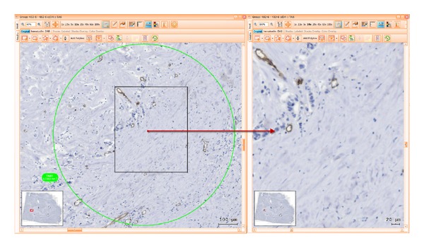
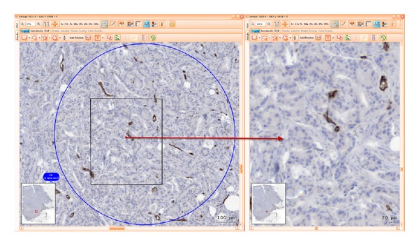
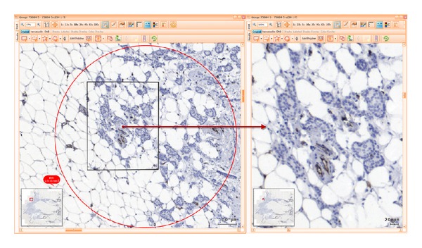
Computerized vessel area measurement was performed using a tissue cytometry system (TissueFAXS) on slides originated from 50 patients with breast cancer. Vessels were marked using immunohistochemistry with CD34. Regions of interest were manually defined for each tumour compartment.
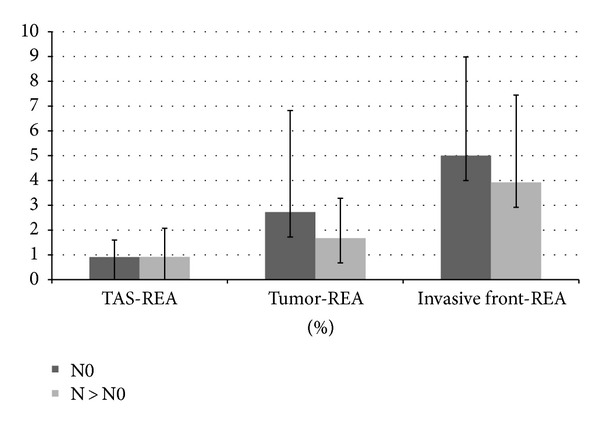
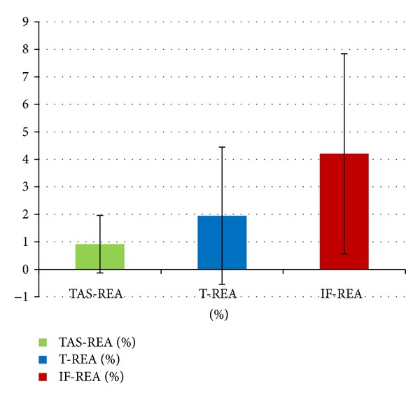
Tumour invasive front vascular endothelia area was 2.15 times higher than that in tumour parenchyma and 4.61 times higher than that in TAS (P < 0.002). Worth to mention that the lymph node negative subgroup of patients show a slight but constant increase of vessel index in all examined compartments of breast tumour.
Whole slide digital examination and region of interest (ROI) analysis are a valuable tool in scoring angiogenesis markers and disclosing their prognostic capacity. Our study reveals compartments' variability of vessel density inside the tumour and highlights the propensity of invasive front to associate an active process of angiogenesis with potential implications in adjuvant therapy.
背景/目的:微血管密度(MVD)定义的肿瘤血管生成通常被认为是乳腺癌的预后因素。然而,由于测量系统和截止值的可变性,到目前为止,它是否有助于预测还存在争议。我们的研究旨在通过比较明确的隔室来对血管异质性进行分级:肿瘤相关基质(TAS)、肿瘤实质和肿瘤浸润前沿。
使用组织细胞计量系统(TissueFAXS)在 50 名乳腺癌患者的幻灯片上进行计算机化血管面积测量。使用 CD34 免疫组织化学标记血管。为每个肿瘤隔室手动定义感兴趣区域。
肿瘤浸润前沿的血管内皮面积是肿瘤实质的 2.15 倍,是 TAS 的 4.61 倍(P<0.002)。值得注意的是,淋巴结阴性亚组的患者在所有检查的乳腺癌肿瘤隔室中,血管指数均略有但持续增加。
全幻灯片数字检查和感兴趣区域(ROI)分析是评分血管生成标志物并揭示其预后能力的有价值工具。我们的研究揭示了肿瘤内血管密度的隔室变异性,并强调了浸润前沿与潜在的血管生成活性过程相关联的倾向,这可能对辅助治疗有影响。