State Key Laboratory of Natural and Biomimetic Drugs, School of Pharmaceutical Sciences, Peking University, Beijing 100191, China.
Part Fibre Toxicol. 2013 Oct 3;10:47. doi: 10.1186/1743-8977-10-47.
Nanocarriers represent an attractive means of drug delivery, but their biosafety must be established before their use in clinical research.
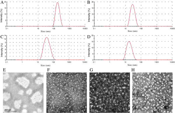
Four kinds of amphiphilic polymeric (PEG-PG-PCL, PEEP-PCL, PEG-PCL and PEG-DSPE) micelles with similar hydrophilic or hydrophobic structure were prepared and their in vitro and in vivo safety were evaluated and compared.
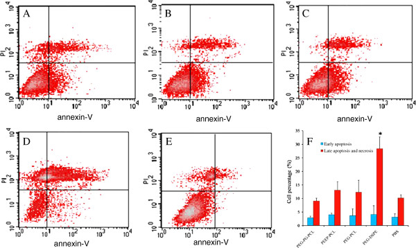
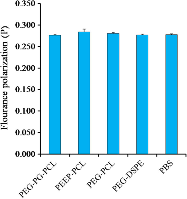
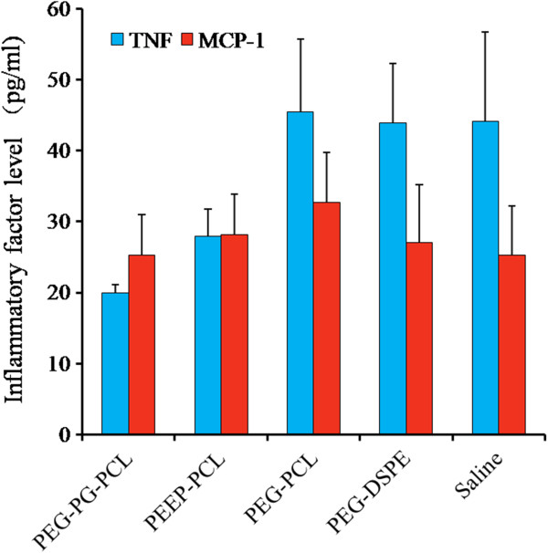
In vitro nanotoxicity evaluations included assessments of cell morphology, cell volume, inflammatory effects, cytotoxicity, apoptosis and membrane fluidity. An umbilical vein cell line (Eahy.926) and a kind of macrophages (J774.A1) were used as cell models considering that intravenous route is dominant for micelle delivery systems. In vivo analyses included complete blood count, lymphocyte subset analysis, detection of plasma inflammatory factors and histological observations of major organs after intravenous administration to KM mice.
All the micelles enhanced inflammatory molecules in J774.A1 cells, likely resulting from the increased ROS levels. PEG-PG-PCL and PEEP-PCL micelles were found to increase the J774.A1 cell volume. This likely correlated with the size of PEG-PG-PCL micelles and the polyphosphoester structure in PEEP-PCL. PEG-DSPE micelles inhibited the growth of Eahy.926 cells via inducing apoptosis. This might relate to the structure of DSPE, which is a type of phospholipid and has good affinity with cell membrane. No evidence was found for cell membrane changes after treatment with these micelles for 24 h. In the in vivo study, during 8 days of 4 time injection, each of the four nanocarriers altered the hematic phase differently without changes in inflammatory factors or pathological changes in target organs.
These results demonstrate that the micelles investigated exhibit diverse nanotoxicity correlated with their structures, their biosafety is different in different cell model, and there is no in vitro and in vivo correlation found. We believe that this study will certainly provide more scientific understandings on the nanotoxicity of amphiphilic polymeric micelles.
纳米载体是一种有吸引力的药物递送方式,但在将其用于临床研究之前,必须确定其生物安全性。
制备四种具有相似亲水或疏水结构的两亲性聚合物(PEG-PG-PCL、PEEP-PCL、PEG-PCL 和 PEG-DSPE)胶束,并对其进行体外和体内安全性评价和比较。
体外纳米毒性评价包括细胞形态、细胞体积、炎症效应、细胞毒性、细胞凋亡和膜流动性评估。考虑到静脉途径是胶束给药系统的主要途径,因此使用脐带静脉细胞系(Eahy.926)和一种巨噬细胞(J774.A1)作为细胞模型。体内分析包括静脉注射后 KM 小鼠的全血细胞计数、淋巴细胞亚群分析、血浆炎症因子检测和主要器官组织学观察。
所有胶束均增强了 J774.A1 细胞中的炎症分子,这可能是由于 ROS 水平升高所致。PEG-PG-PCL 和 PEEP-PCL 胶束被发现增加了 J774.A1 细胞的体积。这可能与 PEG-PG-PCL 胶束的大小和 PEEP-PCL 中的聚磷酸酯结构有关。PEG-DSPE 胶束通过诱导细胞凋亡抑制 Eahy.926 细胞的生长。这可能与 DSPE 的结构有关,DSPE 是一种磷脂,与细胞膜具有良好的亲和力。在用这些胶束处理 24 小时后,没有发现细胞膜发生变化的证据。在体内研究中,在 4 次注射的 8 天期间,四种纳米载体中的每一种都以不同的方式改变了血液相,而炎症因子或靶器官的病理变化没有改变。
这些结果表明,所研究的胶束表现出与结构相关的不同纳米毒性,其生物安全性在不同的细胞模型中不同,并且在体外和体内均未发现相关性。我们相信,这项研究将为两亲性聚合物胶束的纳米毒性提供更科学的认识。