Zhou Tian, Dong Qinglei, Shen Yang, Wu Wei, Wu Haide, Luo Xianglin, Liao Xiaoling, Wang Guixue
Key Laboratory for Biorheological Science and Technology of Ministry of Education, State and Local Joint Engineering Laboratory for Vascular Implants, Bioengineering College of Chongqing University, Chongqing.
Institute of Biomedical Engineering, School of Preclinical and Forensic Medicine, Sichuan University.
Int J Nanomedicine. 2016 Dec 5;11:6517-6531. doi: 10.2147/IJN.S112658. eCollection 2016.
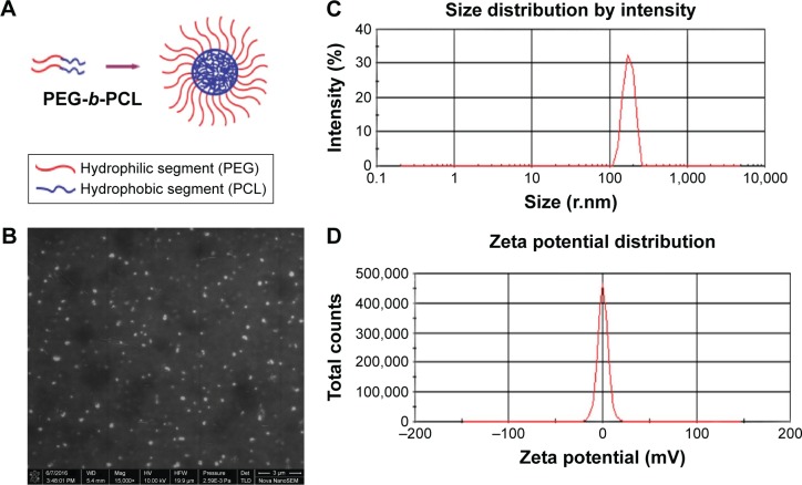
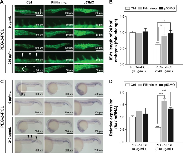
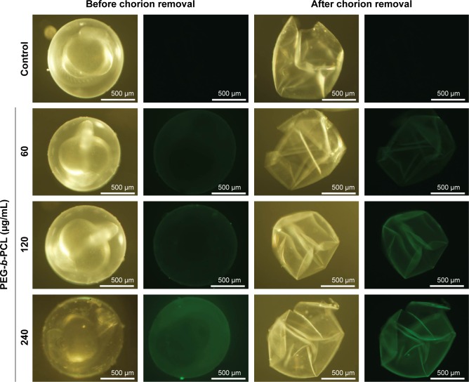
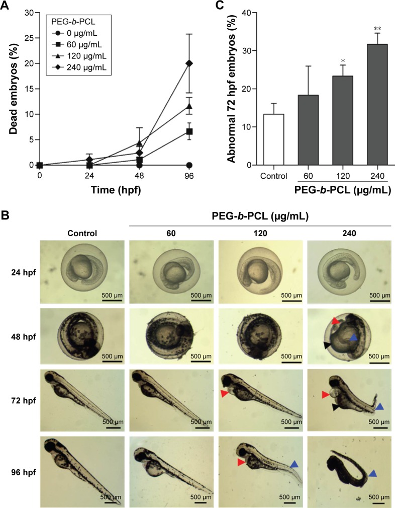
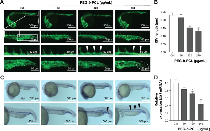
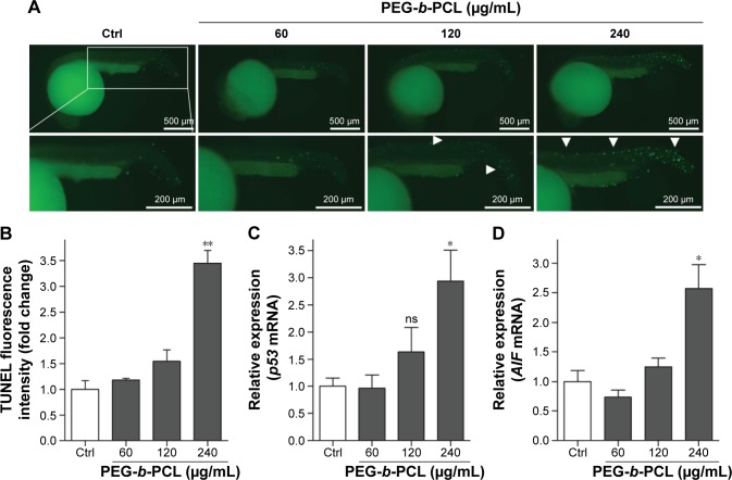
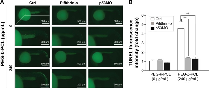
Micro/nanoparticles could cause adverse effects on cardiovascular system and increase the risk for cardiovascular disease-related events. Nanoparticles prepared from poly(ethylene glycol) (PEG)--poly(-caprolactone) (PCL), namely PEG--PCL, a widely studied biodegradable copolymer, are promising carriers for the drug delivery systems. However, it is unknown whether polymeric PEG--PCL nano-micelles give rise to potential complications of the cardiovascular system. Zebrafish were used as an in vivo model to evaluate the effects of PEG--PCL nano-micelle on cardiovascular development. The results showed that PEG--PCL nano-micelle caused embryo mortality as well as embryonic and larval malformations in a dose-dependent manner. To determine PEG--PCL nano-micelle effects on embryonic angiogenesis, a critical process in zebrafish cardiovascular development, growth of intersegmental vessels (ISVs) and caudal vessels (CVs) in flk1-GFP transgenic zebrafish embryos using fluorescent stereomicroscopy were examined. The expression of fetal liver kinase 1 (flk1), an angiogenic factor, by real-time quantitative polymerase chain reaction (qPCR) and in situ whole-mount hybridization were also analyzed. PEG--PCL nano-micelle decreased growth of ISVs and CVs, as well as reduced flk1 expression in a concentration-dependent manner. Parallel to the inhibitory effects on angiogenesis, PEG--PCL nano-micelle exposure upregulated p53 pro-apoptotic pathway and induced cellular apoptosis in angiogenic regions by qPCR and terminal deoxynucleotidyl transferase dUTP nick end labeling (TUNEL) apoptosis assay. This study further showed that inhibiting p53 activity, either by pharmacological inhibitor or RNA interference, could abrogate the apoptosis and angiogenic defects caused by PEG--PCL nano-micelles, indicating that PEG--PCL nano-micelle inhibits angiogenesis by activating p53-mediated apoptosis. This study indicates that polymeric PEG--PCL nano-micelle could pose potential hazards to cardiovascular development.
微米/纳米颗粒可能对心血管系统产生不良影响,并增加心血管疾病相关事件的风险。由聚乙二醇(PEG)-聚(ε-己内酯)(PCL)制备的纳米颗粒,即PEG-PCL,一种被广泛研究的可生物降解共聚物,是药物递送系统中有前景的载体。然而,尚不清楚聚合物PEG-PCL纳米胶束是否会引发心血管系统的潜在并发症。斑马鱼被用作体内模型来评估PEG-PCL纳米胶束对心血管发育的影响。结果表明,PEG-PCL纳米胶束以剂量依赖的方式导致胚胎死亡以及胚胎和幼虫畸形。为了确定PEG-PCL纳米胶束对胚胎血管生成的影响,这是斑马鱼心血管发育中的一个关键过程,使用荧光立体显微镜检查了flk1-GFP转基因斑马鱼胚胎中节间血管(ISV)和尾血管(CV)的生长情况。还通过实时定量聚合酶链反应(qPCR)和原位全胚胎杂交分析了血管生成因子胎儿肝激酶1(flk1)的表达。PEG-PCL纳米胶束以浓度依赖的方式降低了ISV和CV的生长,并降低了flk1的表达。与对血管生成的抑制作用平行,PEG-PCL纳米胶束暴露上调了p53促凋亡途径,并通过qPCR和末端脱氧核苷酸转移酶dUTP缺口末端标记(TUNEL)凋亡检测在血管生成区域诱导细胞凋亡。这项研究进一步表明,通过药物抑制剂或RNA干扰抑制p53活性,可以消除PEG-PCL纳米胶束引起的凋亡和血管生成缺陷,表明PEG-PCL纳米胶束通过激活p53介导的凋亡来抑制血管生成。这项研究表明,聚合物PEG-PCL纳米胶束可能对心血管发育构成潜在危害。