• 文献检索
  • 文档翻译
  • 深度研究
  • 学术资讯
  • Suppr Zotero 插件Zotero 插件
  • 邀请有礼
  • 套餐&价格
  • 历史记录
应用&插件
Suppr Zotero 插件Zotero 插件浏览器插件Mac 客户端Windows 客户端微信小程序
定价
高级版会员购买积分包购买API积分包
服务
文献检索文档翻译深度研究API 文档MCP 服务
关于我们
关于 Suppr公司介绍联系我们用户协议隐私条款
关注我们

Suppr 超能文献

核心技术专利:CN118964589B侵权必究
粤ICP备2023148730 号-1Suppr @ 2026

文献检索

告别复杂PubMed语法,用中文像聊天一样搜索,搜遍4000万医学文献。AI智能推荐,让科研检索更轻松。

立即免费搜索

文件翻译

保留排版,准确专业,支持PDF/Word/PPT等文件格式,支持 12+语言互译。

免费翻译文档

深度研究

AI帮你快速写综述,25分钟生成高质量综述,智能提取关键信息,辅助科研写作。

立即免费体验

非晶态二氧化硅纳米颗粒诱导 L-02 人肝细胞系的多核化和细胞功能障碍。

Multinucleation and cell dysfunction induced by amorphous silica nanoparticles in an L-02 human hepatic cell line.

机构信息

School of Public Health, Capital Medical University, Beijing ; Beijing Key Laboratory of Environmental Toxicology, Capital Medical University, Beijing ; School of Public Health, Jilin University, Changchun, Jilin.

出版信息

Int J Nanomedicine. 2013;8:3533-41. doi: 10.2147/IJN.S46732. Epub 2013 Sep 19.

DOI:10.2147/IJN.S46732
PMID:24092974
原文链接:https://pmc.ncbi.nlm.nih.gov/articles/PMC3787934/
Abstract

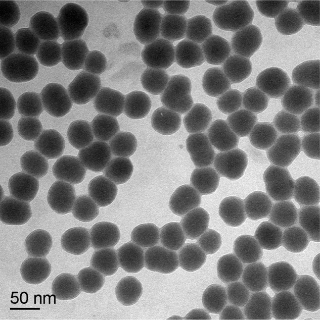
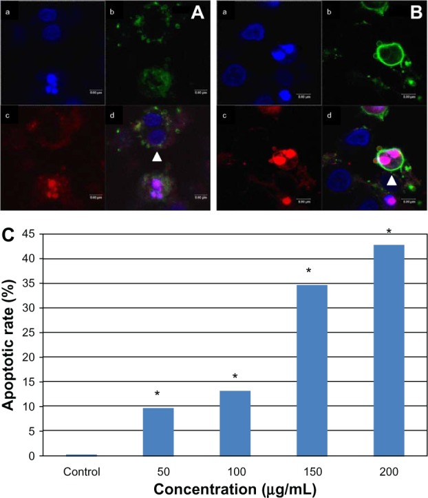
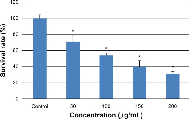
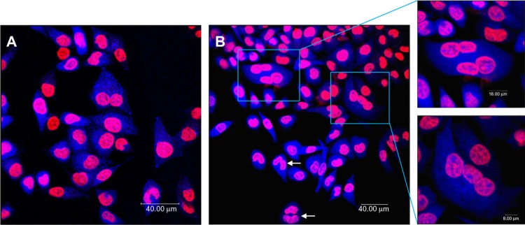
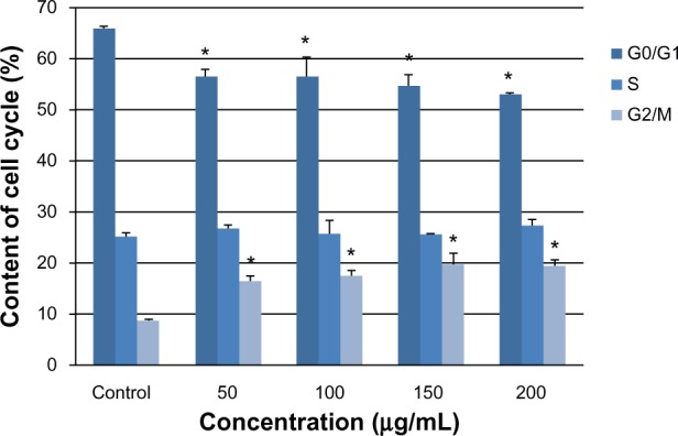
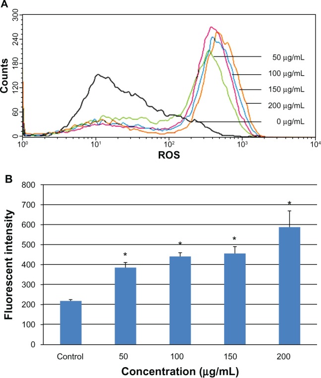
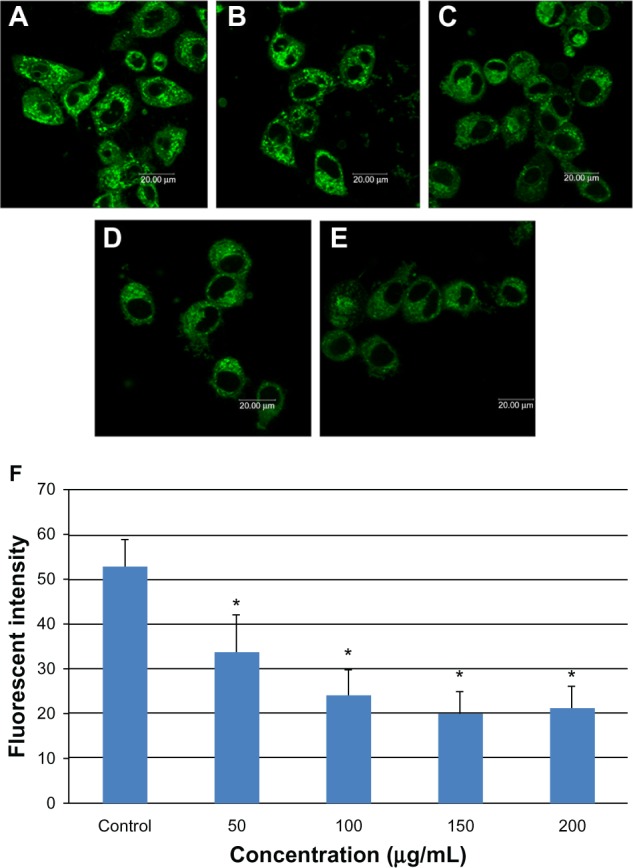
Silica nanoparticles (SNPs) are one of the most important nanomaterials, and have been widely used in a variety of fields. Therefore, their effects on human health and the environment have been addressed in a number of studies. In this work, the effects of amorphous SNPs were investigated with regard to multinucleation in L-02 human hepatic cells. Our results show that L-02 cells had an abnormally high incidence of multinucleation upon exposure to silica, that increased in a dose-dependent manner. Propidium iodide staining showed that multinucleated cells were arrested in G2/M phase of the cell cycle. Increased multinucleation in L-02 cells was associated with increased generation of cellular reactive oxygen species and mitochondrial damage on flow cytometry and confocal microscopy, which might have led to failure of cytokinesis in these cells. Further, SNPs inhibited cell growth and induced apoptosis in exposed cells. Taken together, our findings demonstrate that multinucleation in L-02 human hepatic cells might be a failure to undergo cytokinesis or cell fusion in response to SNPs, and the increase in cellular reactive oxygen species could be responsible for the apoptosis seen in both mononuclear cells and multinucleated cells.

摘要

硅纳米颗粒(SNPs)是最重要的纳米材料之一,已广泛应用于许多领域。因此,许多研究都探讨了 SNPs 对人类健康和环境的影响。在这项工作中,研究了非晶态 SNPs 对 L-02 人肝细胞多核化的影响。我们的结果表明,暴露于硅后,L-02 细胞的多核化异常高,且呈剂量依赖性增加。碘化丙啶染色表明,多核细胞在细胞周期的 G2/M 期被阻滞。流式细胞术和共聚焦显微镜显示,L-02 细胞中多核化的增加与细胞内活性氧(ROS)的产生增加和线粒体损伤有关,这可能导致这些细胞的胞质分裂失败。此外,SNPs 抑制暴露细胞的生长并诱导细胞凋亡。总之,我们的研究结果表明,L-02 人肝细胞的多核化可能是对 SNPs 反应时未能进行胞质分裂或细胞融合,细胞内活性氧(ROS)的增加可能是单核细胞和多核细胞凋亡的原因。

https://cdn.ncbi.nlm.nih.gov/pmc/blobs/cf49/3787934/9c23d47378e5/ijn-8-3533Fig8.jpg
https://cdn.ncbi.nlm.nih.gov/pmc/blobs/cf49/3787934/0d75da984449/ijn-8-3533Fig1.jpg
https://cdn.ncbi.nlm.nih.gov/pmc/blobs/cf49/3787934/3601e4c05289/ijn-8-3533Fig2.jpg
https://cdn.ncbi.nlm.nih.gov/pmc/blobs/cf49/3787934/c8e678eebc48/ijn-8-3533Fig3.jpg
https://cdn.ncbi.nlm.nih.gov/pmc/blobs/cf49/3787934/41be03b45edc/ijn-8-3533Fig4.jpg
https://cdn.ncbi.nlm.nih.gov/pmc/blobs/cf49/3787934/24981c995e32/ijn-8-3533Fig5.jpg
https://cdn.ncbi.nlm.nih.gov/pmc/blobs/cf49/3787934/89cafa0eb7f9/ijn-8-3533Fig6.jpg
https://cdn.ncbi.nlm.nih.gov/pmc/blobs/cf49/3787934/488ff3ae887f/ijn-8-3533Fig7.jpg
https://cdn.ncbi.nlm.nih.gov/pmc/blobs/cf49/3787934/9c23d47378e5/ijn-8-3533Fig8.jpg

相似文献

1
Multinucleation and cell dysfunction induced by amorphous silica nanoparticles in an L-02 human hepatic cell line.非晶态二氧化硅纳米颗粒诱导 L-02 人肝细胞系的多核化和细胞功能障碍。
Int J Nanomedicine. 2013;8:3533-41. doi: 10.2147/IJN.S46732. Epub 2013 Sep 19.
2
Amorphous silica nanoparticles cause abnormal cytokinesis and multinucleation through dysfunction of the centralspindlin complex and microfilaments.无定形二氧化硅纳米颗粒通过中央纺锤体复合物和微丝的功能障碍引起异常的胞质分裂和多核化。
Part Fibre Toxicol. 2023 Aug 22;20(1):34. doi: 10.1186/s12989-023-00544-8.
3
In vitro cytotoxicity and induction of apoptosis by silica nanoparticles in human HepG2 hepatoma cells.体外实验中二氧化硅纳米颗粒对人肝癌 HepG2 细胞的细胞毒性和诱导凋亡作用。
Int J Nanomedicine. 2011;6:1889-901. doi: 10.2147/IJN.S24005. Epub 2011 Sep 7.
4
Aberrant cytokinesis and cell fusion result in multinucleation in HepG2 cells exposed to silica nanoparticles.异常的胞质分裂和细胞融合导致暴露于二氧化硅纳米颗粒的HepG2细胞出现多核现象。
Chem Res Toxicol. 2015 Mar 16;28(3):490-500. doi: 10.1021/tx500473h. Epub 2015 Jan 27.
5
Impact of silver nanoparticles on human cells: effect of particle size.纳米银颗粒对人体细胞的影响:粒径的影响。
Nanotoxicology. 2010 Sep;4(3):319-30. doi: 10.3109/17435390.2010.483745.
6
Silver Nanoparticles Exhibit the Dose-Dependent Anti-Proliferative Effect against Human Squamous Carcinoma Cells Attenuated in the Presence of Berberine.银纳米颗粒对人鳞状癌细胞具有剂量依赖性抗增殖作用,在小檗碱存在下这种作用减弱。
Molecules. 2016 Mar 17;21(3):365. doi: 10.3390/molecules21030365.
7
Cytotoxicity and apoptosis induced by silver nanoparticles in human liver HepG2 cells in different dispersion media.不同分散介质中银纳米颗粒对人肝癌HepG2细胞的细胞毒性和凋亡诱导作用。
J Appl Toxicol. 2016 Mar;36(3):352-60. doi: 10.1002/jat.3199. Epub 2015 Jul 21.
8
Size-dependent cytotoxicity of amorphous silica nanoparticles in human hepatoma HepG2 cells.粒径依赖的无定形二氧化硅纳米颗粒对人肝癌 HepG2 细胞的细胞毒性。
Toxicol In Vitro. 2011 Oct;25(7):1343-52. doi: 10.1016/j.tiv.2011.05.003. Epub 2011 May 7.
9
Cytotoxicity and genotoxicity of silver nanoparticles in human cells.银纳米颗粒对人体细胞的细胞毒性和遗传毒性。
ACS Nano. 2009 Feb 24;3(2):279-90. doi: 10.1021/nn800596w.
10
Multi-platform genotoxicity analysis of silver nanoparticles in the model cell line CHO-K1.多平台遗传毒性分析银纳米粒子在模式细胞系 CHO-K1。
Toxicol Lett. 2013 Sep 12;222(1):55-63. doi: 10.1016/j.toxlet.2013.07.011. Epub 2013 Jul 18.

引用本文的文献

1
Fine particulate matter‑induced cardiac developmental toxicity (Review).细颗粒物诱发的心脏发育毒性(综述)
Exp Ther Med. 2024 Oct 29;29(1):6. doi: 10.3892/etm.2024.12756. eCollection 2025 Jan.
2
Multinuclei Occurred Under Cryopreservation and Enhanced the Pathogenicity of .多核现象在冷冻保存过程中出现并增强了……的致病性。 (原文中“Enhanced the Pathogenicity of.”后面似乎缺少具体内容)
Front Microbiol. 2021 Jun 25;12:650902. doi: 10.3389/fmicb.2021.650902. eCollection 2021.
3
PARP-1 overexpression does not protect HaCaT cells from DNA damage induced by SiO nanoparticles.

本文引用的文献

1
Mechanism of cellular uptake of genotoxic silica nanoparticles.细胞摄取遗传毒性二氧化硅纳米颗粒的机制。
Part Fibre Toxicol. 2012 Jul 23;9:29. doi: 10.1186/1743-8977-9-29.
2
Frequent aneuploidy among normal human hepatocytes.正常人类肝细胞中频繁的非整倍体。
Gastroenterology. 2012 Jan;142(1):25-8. doi: 10.1053/j.gastro.2011.10.029. Epub 2011 Nov 2.
3
The cytotoxic activity of amorphous silica nanoparticles is mainly influenced by surface area and not by aggregation.无定形二氧化硅纳米颗粒的细胞毒性活性主要受表面积影响,而不受聚集影响。
聚(ADP-核糖)聚合酶-1(PARP-1)的过表达并不能保护角质形成细胞(HaCaT细胞)免受二氧化硅纳米颗粒诱导的DNA损伤。
Toxicol Res (Camb). 2021 Apr 12;10(3):399-408. doi: 10.1093/toxres/tfaa110. eCollection 2021 May.
4
Quantitative evaluation of cellular internalization of polymeric nanoparticles within laryngeal cancer cells and immune cells for enhanced drug delivery.聚合物纳米颗粒在喉癌细胞和免疫细胞内摄取的定量评估以增强药物递送
Nanoscale Res Lett. 2021 Mar 2;16(1):40. doi: 10.1186/s11671-021-03498-y.
5
The chronic effect of amorphous silica nanoparticles and benzo[]pyrene co-exposure at low dose in human bronchial epithelial BEAS-2B cells.低剂量无定形二氧化硅纳米颗粒与苯并[a]芘共同暴露对人支气管上皮BEAS-2B细胞的慢性影响。
Toxicol Res (Camb). 2019 Aug 12;8(5):731-740. doi: 10.1039/c9tx00112c. eCollection 2019 Sep 1.
6
Toxicology of silica nanoparticles: an update.硅纳米粒子的毒理学:最新进展。
Arch Toxicol. 2017 Sep;91(9):2967-3010. doi: 10.1007/s00204-017-1993-y. Epub 2017 Jun 1.
7
Co-exposure to amorphous silica nanoparticles and benzo[a]pyrene at low level in human bronchial epithelial BEAS-2B cells.人支气管上皮 BEAS-2B 细胞中低水平的无定形二氧化硅纳米颗粒和苯并[a]芘的共暴露。
Environ Sci Pollut Res Int. 2016 Nov;23(22):23134-23144. doi: 10.1007/s11356-016-7559-3. Epub 2016 Sep 3.
Toxicol Lett. 2011 Oct 10;206(2):197-203. doi: 10.1016/j.toxlet.2011.07.013. Epub 2011 Jul 22.
4
Activation of stress-related signalling pathway in human cells upon SiO2 nanoparticles exposure as an early indicator of cytotoxicity.二氧化硅纳米颗粒暴露后人类细胞应激相关信号通路的激活作为细胞毒性的早期标志物。
J Nanobiotechnology. 2011 Jul 29;9:29. doi: 10.1186/1477-3155-9-29.
5
Cytotoxicity and mitochondrial damage caused by silica nanoparticles.硅纳米颗粒导致的细胞毒性和线粒体损伤。
Toxicol In Vitro. 2011 Dec;25(8):1619-29. doi: 10.1016/j.tiv.2011.06.012. Epub 2011 Jun 24.
6
Size-dependent cytotoxicity of amorphous silica nanoparticles in human hepatoma HepG2 cells.粒径依赖的无定形二氧化硅纳米颗粒对人肝癌 HepG2 细胞的细胞毒性。
Toxicol In Vitro. 2011 Oct;25(7):1343-52. doi: 10.1016/j.tiv.2011.05.003. Epub 2011 May 7.
7
Proteomic characterization of engineered nanomaterial-protein interactions in relation to surface reactivity.工程纳米材料-蛋白质相互作用的蛋白质组学特征与表面反应性有关。
ACS Nano. 2011 Jun 28;5(6):4300-9. doi: 10.1021/nn101492k. Epub 2011 May 4.
8
Inflammatory and cytotoxic responses of an alveolar-capillary coculture model to silica nanoparticles: comparison with conventional monocultures.肺泡毛细血管共培养模型对二氧化硅纳米颗粒的炎症和细胞毒性反应:与传统的单层细胞培养比较。
Part Fibre Toxicol. 2011 Jan 27;8(1):6. doi: 10.1186/1743-8977-8-6.
9
Amorphous nanosilica induce endocytosis-dependent ROS generation and DNA damage in human keratinocytes.无定形纳米二氧化硅诱导人角质形成细胞内吞依赖性 ROS 生成和 DNA 损伤。
Part Fibre Toxicol. 2011 Jan 15;8:1. doi: 10.1186/1743-8977-8-1.
10
The nanosilica hazard: another variable entity.纳米二氧化硅的危害:另一个可变实体。
Part Fibre Toxicol. 2010 Dec 3;7(1):39. doi: 10.1186/1743-8977-7-39.