Centre for Molecular NanoScience (CMNS), University of Leeds, Leeds LS2 9JT, UK.
Part Fibre Toxicol. 2012 Jul 23;9:29. doi: 10.1186/1743-8977-9-29.
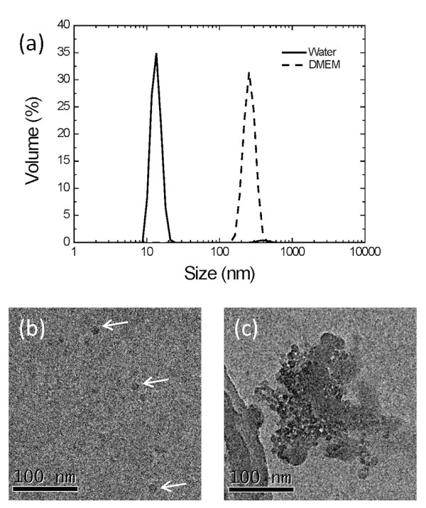
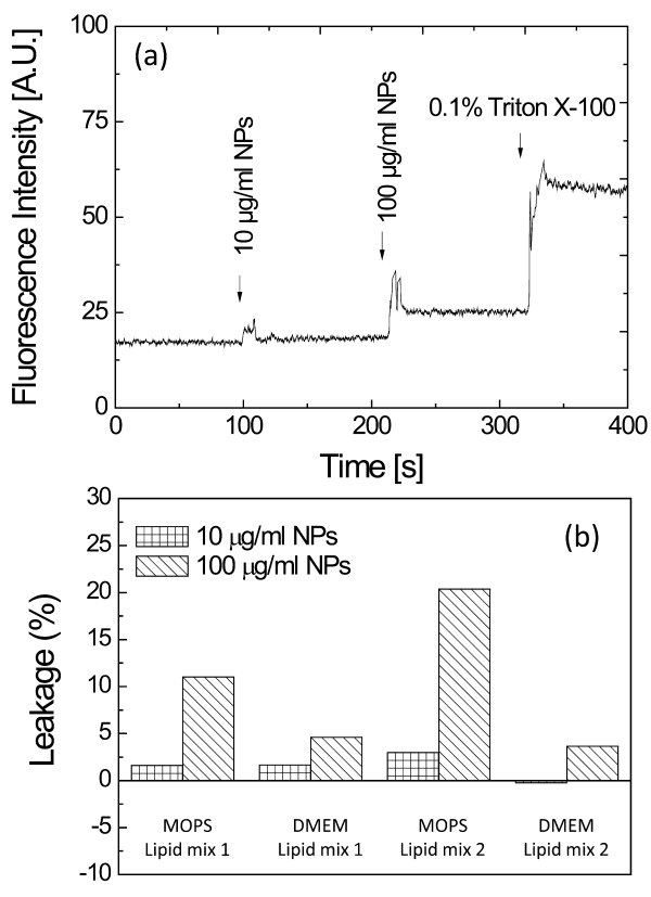
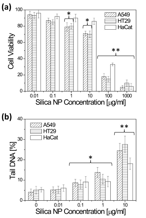
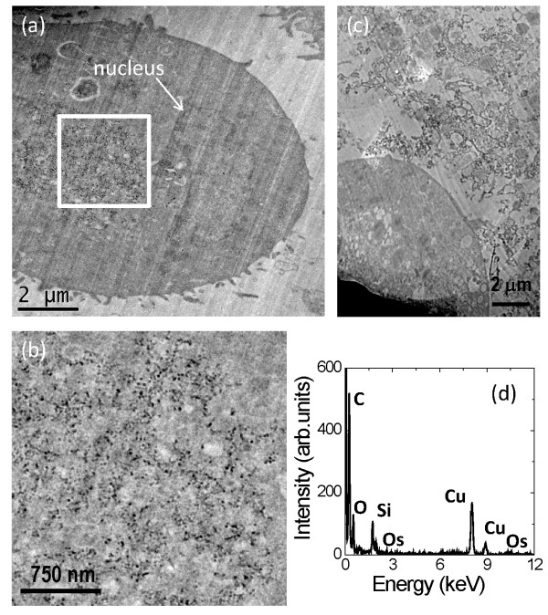
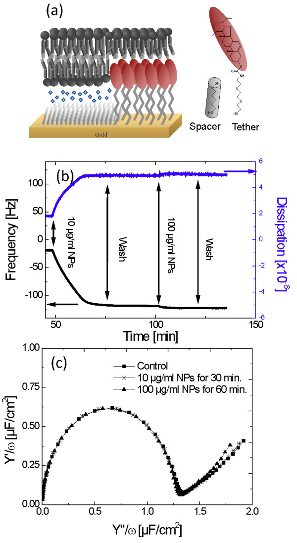
Mechanisms for cellular uptake of nanoparticles have important implications for nanoparticulate drug delivery and toxicity. We have explored the mechanism of uptake of amorphous silica nanoparticles of 14 nm diameter, which agglomerate in culture medium to hydrodynamic diameters around 500 nm. In HT29, HaCat and A549 cells, cytotoxicity was observed at nanoparticle concentrations ≥ 1 μg/ml, but DNA damage was evident at 0.1 μg/ml and above. Transmission electron microscopy (TEM) combined with energy-dispersive X-ray spectroscopy confirmed entry of the silica particles into A549 cells exposed to 10 μg/ml of nanoparticles. The particles were observed in the cytoplasm but not within membrane bound vesicles or in the nucleus. TEM of cells exposed to nanoparticles at 4°C for 30 minutes showed particles enter cells when activity is low, suggesting a passive mode of entry. Plasma lipid membrane models identified physical interactions between the membrane and the silica NPs. Quartz crystal microbalance experiments on tethered bilayer lipid membrane systems show that the nanoparticles strongly bind to lipid membranes, forming an adherent monolayer on the membrane. Leakage assays on large unilamellar vesicles (400 nm diameter) indicate that binding of the silica NPs transiently disrupts the vesicles which rapidly self-seal. We suggest that an adhesive interaction between silica nanoparticles and lipid membranes could cause passive cellular uptake of the particles.
纳米颗粒的细胞摄取机制对纳米颗粒药物传递和毒性具有重要意义。我们研究了直径为 14nm 的无定形二氧化硅纳米颗粒的摄取机制,这些颗粒在培养基中聚集,水动力直径约为 500nm。在 HT29、HaCat 和 A549 细胞中,在纳米颗粒浓度≥1μg/ml 时观察到细胞毒性,但在 0.1μg/ml 及以上时明显出现 DNA 损伤。透射电子显微镜(TEM)结合能量色散 X 射线能谱证实,暴露于 10μg/ml 纳米颗粒的 A549 细胞中有二氧化硅颗粒进入。这些颗粒在细胞质中观察到,但不在膜结合囊泡内或核内。在 4°C 下将细胞暴露于纳米颗粒 30 分钟的 TEM 显示,当活性较低时,颗粒进入细胞,表明进入细胞的方式为被动模式。等离子脂质膜模型确定了膜与二氧化硅 NPs 之间的物理相互作用。在连接双层脂质膜系统上的石英晶体微天平实验表明,纳米颗粒强烈结合到脂质膜上,在膜上形成附着的单层。对大单层囊泡(400nm 直径)的泄漏实验表明,二氧化硅纳米颗粒的结合会短暂破坏囊泡,囊泡会迅速自我密封。我们认为,二氧化硅纳米颗粒与脂质膜之间的粘附相互作用可能导致颗粒的被动细胞摄取。