Department of Biomedical Engineering, Wayne State University, 2322 Engineering, 5050 Anthony Wayne Dr,, Detroit, MI 48202, USA.
Cardiovasc Diabetol. 2013 Oct 5;12:142. doi: 10.1186/1475-2840-12-142.
Endothelial dysfunction precedes pathogenesis of vascular complications in diabetes. In recent years, the mechanisms of endothelial dysfunction were investigated to outline strategies for its treatment. However, the therapies for dysfunctional endothelium resulted in multiple clinical trial failures and remain elusive. There is a need for defining hyperglycemia-induced endothelial dysfunction with both generic and specific dysfunctional changes in endothelial cells (EC) using a systems approach. In this study, we investigated hyperglycemia-induced endothelial dysfunction in HUVEC and HMVEC. We investigated hyperglycemia-induced functional changes (superoxide (O₂⁻), and hydrogen peroxide (H₂O₂) production and mitochondrial membrane polarization) and gene expression fingerprints of related enzymes (nitric oxide synthase, NAD(P)H oxidase, and reactive oxygen species (ROS) neutralizing enzymes) in both ECs.
Gene expression of NOS2, NOS3, NOX4, CYBA, UCP1, CAT, TXNRD1, TXNRD2, GPX1, NOX1, SOD1, SOD2, PRDX1, 18s, and RPLP0 were measured using real-time PCR. O₂⁻ production was measured with dihydroethidium (DHE) fluorescence measurement. H2O2 production was measured using Amplex Red assay. Mitochondrial membrane polarization was measured using JC-10 based fluorescence measurement.
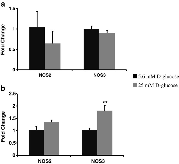
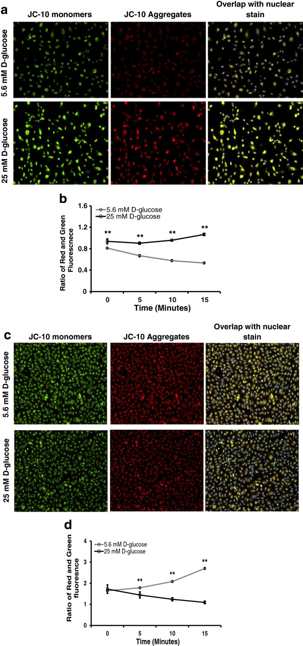
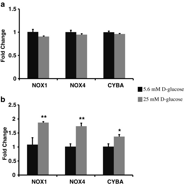
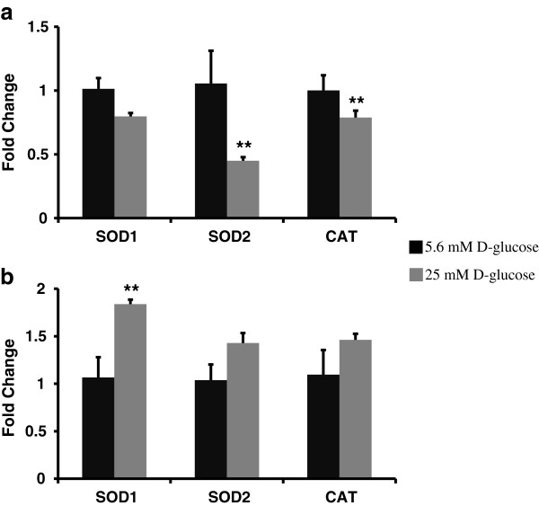
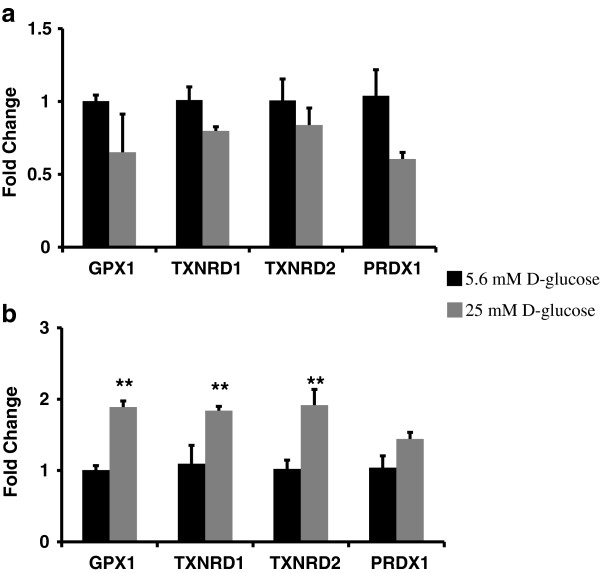
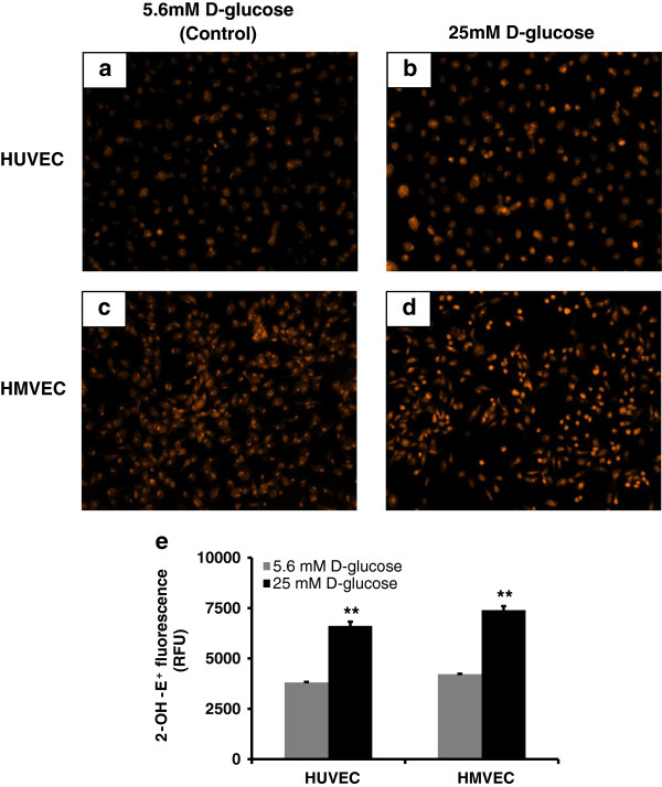
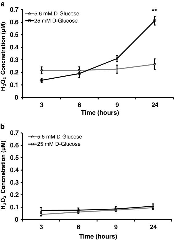
We showed that the O₂⁻ levels increased similarly in both ECs with hyperglycemia. However, these endothelial cells showed significantly different underlying gene expression profile, H₂O₂ production and mitochondrial membrane polarization. In HUVEC, hyperglycemia increased H₂O₂ production, and hyperpolarized mitochondrial membrane. ROS neutralizing enzymes SOD2 and CAT gene expression were downregulated. In contrast, there was an upregulation of nitric oxide synthase and NAD(P)H oxidase and a depolarization of mitochondrial membrane in HMVEC. In addition, ROS neutralizing enzymes SOD1, GPX1, TXNRD1 and TXNRD2 gene expression were significantly upregulated in high glucose treated HMVEC.
Our findings highlighted a unique framework for hyperglycemia-induced endothelial dysfunction. We showed that multiple pathways are differentially affected in these endothelial cells in hyperglycemia. High occurrences of gene expression changes in HMVEC in this study supports the hypothesis that microvasculature precedes macrovasculature in epigenetic regulation forming vascular metabolic memory. Identifying genomic phenotype and corresponding functional changes in hyperglycemic endothelial dysfunction will provide a suitable systems biology approach for understanding underlying mechanisms and possible effective therapeutic intervention.
血管并发症的发病机制先于内皮功能障碍在糖尿病中出现。近年来,人们对内皮功能障碍的机制进行了研究,以制定治疗策略。然而,针对功能失调内皮细胞的治疗方法在多项临床试验中均以失败告终,至今仍未找到有效的方法。因此,我们需要使用系统方法来定义高血糖引起的内皮细胞的一般和特定功能障碍变化。在这项研究中,我们研究了高血糖诱导的 HUVEC 和 HMVEC 中的内皮功能障碍。我们研究了高血糖诱导的功能变化(超氧化物 (O₂⁻) 和过氧化氢 (H₂O₂) 的产生以及线粒体膜的极化)以及相关酶(一氧化氮合酶、NAD(P)H 氧化酶和活性氧 (ROS) 中和酶)的基因表达特征。
使用实时 PCR 测量 NOS2、NOS3、NOX4、CYBA、UCP1、CAT、TXNRD1、TXNRD2、GPX1、NOX1、SOD1、SOD2、PRDX1、18s 和 RPLP0 的基因表达。使用二氢乙锭 (DHE) 荧光测量法测量 O₂⁻的产生。使用 Amplex Red 测定法测量 H₂O₂的产生。使用基于 JC-10 的荧光测量法测量线粒体膜的极化。
我们表明,高血糖会导致两种内皮细胞的 O₂⁻水平相似增加。然而,这些内皮细胞表现出明显不同的潜在基因表达谱、H₂O₂产生和线粒体膜极化。在 HUVEC 中,高血糖会增加 H₂O₂的产生,并使线粒体膜超极化。ROS 中和酶 SOD2 和 CAT 的基因表达下调。相比之下,HMVEC 中的一氧化氮合酶和 NAD(P)H 氧化酶上调,线粒体膜去极化。此外,高葡萄糖处理的 HMVEC 中 ROS 中和酶 SOD1、GPX1、TXNRD1 和 TXNRD2 的基因表达显著上调。
我们的研究结果突出了一种独特的高血糖诱导内皮功能障碍的框架。我们表明,这些内皮细胞中存在多种通路受到高血糖的不同影响。本研究中 HMVEC 中大量基因表达变化的发生支持这样一种假设,即血管代谢记忆中,微血管先于大血管在表观遗传调节中形成。确定高血糖性内皮功能障碍的基因组表型和相应的功能变化将为理解潜在机制和可能的有效治疗干预提供合适的系统生物学方法。