• 文献检索
  • 文档翻译
  • 深度研究
  • 学术资讯
  • Suppr Zotero 插件Zotero 插件
  • 邀请有礼
  • 套餐&价格
  • 历史记录
应用&插件
Suppr Zotero 插件Zotero 插件浏览器插件Mac 客户端Windows 客户端微信小程序
定价
高级版会员购买积分包购买API积分包
服务
文献检索文档翻译深度研究API 文档MCP 服务
关于我们
关于 Suppr公司介绍联系我们用户协议隐私条款
关注我们

Suppr 超能文献

核心技术专利:CN118964589B侵权必究
粤ICP备2023148730 号-1Suppr @ 2026

文献检索

告别复杂PubMed语法,用中文像聊天一样搜索,搜遍4000万医学文献。AI智能推荐,让科研检索更轻松。

立即免费搜索

文件翻译

保留排版,准确专业,支持PDF/Word/PPT等文件格式,支持 12+语言互译。

免费翻译文档

深度研究

AI帮你快速写综述,25分钟生成高质量综述,智能提取关键信息,辅助科研写作。

立即免费体验

相似文献

1
Silver nanowire exposure results in internalization and toxicity to Daphnia magna.银纳米线暴露会导致大型溞的内化和毒性。
ACS Nano. 2013 Dec 23;7(12):10681-94. doi: 10.1021/nn4034103. Epub 2013 Dec 5.
2
Aquatic Toxicity Comparison of Silver Nanoparticles and Silver Nanowires.银纳米颗粒与银纳米线的水生毒性比较
Biomed Res Int. 2015;2015:893049. doi: 10.1155/2015/893049. Epub 2015 Jun 1.
3
Dimension-dependent toxicity of silver nanomaterials on the cladocerans Daphnia magna and Daphnia galeata.银纳米材料对大型溞和盔形溞的尺寸依赖性毒性
Chemosphere. 2017 Oct;185:205-212. doi: 10.1016/j.chemosphere.2017.07.011. Epub 2017 Jul 5.
4
Silver nanoparticle toxicity to Daphnia magna is a function of dissolved silver concentration.银纳米颗粒对大型溞的毒性是溶解态银浓度的函数。
Environ Toxicol Chem. 2013 Oct;32(10):2356-64. doi: 10.1002/etc.2300. Epub 2013 Aug 9.
5
Toxicity of two types of silver nanoparticles to aquatic crustaceans Daphnia magna and Thamnocephalus platyurus.两种类型的银纳米粒子对水生甲壳类动物大型溞和扁蜉的毒性。
Environ Sci Pollut Res Int. 2013 May;20(5):3456-63. doi: 10.1007/s11356-012-1290-5. Epub 2012 Nov 11.
6
Sunlight-driven reduction of silver ion to silver nanoparticle by organic matter mitigates the acute toxicity of silver to Daphnia magna.阳光驱动的有机物将银离子还原为银纳米颗粒,减轻了银对大型溞的急性毒性。
J Environ Sci (China). 2015 Sep 1;35:62-68. doi: 10.1016/j.jes.2015.03.007. Epub 2015 May 28.
7
Comparison of nanosilver and ionic silver toxicity in Daphnia magna and Pimephales promelas.纳米银和离子银对大型溞和蓝鳃太阳鱼的毒性比较。
Environ Toxicol Chem. 2012 Nov;31(11):2557-63. doi: 10.1002/etc.1978. Epub 2012 Sep 20.
8
Exposure medium: key in identifying free Ag+ as the exclusive species of silver nanoparticles with acute toxicity to Daphnia magna.暴露介质:确定游离Ag⁺ 是对大型溞具有急性毒性的银纳米颗粒的唯一物种的关键。
Sci Rep. 2015 Apr 10;5:9674. doi: 10.1038/srep09674.
9
Effects of silver nanoparticle properties, media pH and dissolved organic matter on toxicity to Daphnia magna.银纳米颗粒特性、介质pH值和溶解有机物对大型溞毒性的影响。
Ecotoxicol Environ Saf. 2015 Jan;111:263-70. doi: 10.1016/j.ecoenv.2014.09.031. Epub 2014 Nov 5.
10
Importance of surface coatings and soluble silver in silver nanoparticles toxicity to Daphnia magna.表面涂层和可溶性银在银纳米颗粒对大型溞毒性中的重要性。
Nanotoxicology. 2012 Jun;6(4):361-70. doi: 10.3109/17435390.2011.579632. Epub 2011 May 18.

引用本文的文献

1
Last Fifteen Years of Nanotechnology Application with Our Contribute.在我们的贡献下纳米技术应用的过去十五年。
Nanomaterials (Basel). 2025 Feb 10;15(4):265. doi: 10.3390/nano15040265.
2
Cytotoxicity and Genotoxicity Effects of a Magnetic Zeolite Composite in (Straus, 1820).磁性沸石复合材料对 (Straus,1820)的细胞毒性和遗传毒性作用。
Int J Mol Sci. 2024 Jul 9;25(14):7542. doi: 10.3390/ijms25147542.
3
Daphnia magna as a Model Organism to Predict the Teratogenic Effect of Different Compounds.大型溞作为一种模式生物预测不同化合物的致畸作用。
Methods Mol Biol. 2024;2753:261-281. doi: 10.1007/978-1-0716-3625-1_13.
4
Influence of Silver Fiber Morphology on the Dose-Response Relationship and Enrichment in Studied by Elemental Imaging with LA-ICP-TOF-MS.利用 LA-ICP-TOF-MS 元素成像研究银纤维形态对剂量-反应关系和富集的影响。
Chem Res Toxicol. 2024 Feb 19;37(2):292-301. doi: 10.1021/acs.chemrestox.3c00293. Epub 2024 Jan 8.
5
Building the Bridge From Aquatic Nanotoxicology to Safety by Design Silver Nanoparticles.构建从水生纳米毒理学通向设计型银纳米颗粒安全性的桥梁。
Front Bioeng Biotechnol. 2022 Mar 8;10:836742. doi: 10.3389/fbioe.2022.836742. eCollection 2022.
6
Determination of metallic nanoparticles in biological samples by single particle ICP-MS: a systematic review from sample collection to analysis.通过单颗粒电感耦合等离子体质谱法测定生物样品中的金属纳米颗粒:从样品采集到分析的系统综述
Environ Sci Nano. 2022 Jan 13;9(2):420-453. doi: 10.1039/d1en00680k. eCollection 2022 Feb 17.
7
U.S. Federal Agency interests and key considerations for new approach methodologies for nanomaterials.美国联邦机构对纳米材料新方法的兴趣和主要考虑因素。
ALTEX. 2022;39(2):183–206. doi: 10.14573/altex.2105041. Epub 2021 Dec 3.
8
Modeling the metabolic profile of Mytilus edulis reveals molecular signatures linked to gonadal development, sex and environmental site.模拟贻贝的代谢特征揭示了与性腺发育、性别和环境地点相关的分子特征。
Sci Rep. 2021 Jun 18;11(1):12882. doi: 10.1038/s41598-021-90494-y.
9
Crumpling of silver nanowires by endolysosomes strongly reduces toxicity.内溶酶体的银纳米线皱缩强烈降低了毒性。
Proc Natl Acad Sci U S A. 2019 Jul 23;116(30):14893-14898. doi: 10.1073/pnas.1820041116. Epub 2019 Jul 8.
10
Fate and toxicity of silver nanoparticles in freshwater from laboratory to realistic environments: a review.银纳米颗粒在淡水环境中的命运和毒性:从实验室到现实环境的综述。
Environ Sci Pollut Res Int. 2019 Mar;26(8):7390-7404. doi: 10.1007/s11356-019-04150-0. Epub 2019 Jan 23.

本文引用的文献

1
Silver release from silver nanoparticles in natural waters.天然水中纳米银的银释放。
Environ Sci Technol. 2013 May 7;47(9):4140-6. doi: 10.1021/es304023p. Epub 2013 Apr 8.
2
Pattern transfer of aligned metal nano/microwires as flexible transparent electrodes using an electrospun nanofiber template.采用静电纺丝纳米纤维模板实现金属纳米/微米线的对齐转移,用作柔性透明电极。
Nanotechnology. 2013 Feb 8;24(5):055301. doi: 10.1088/0957-4484/24/5/055301. Epub 2013 Jan 11.
3
Use of silver nanowires to determine thresholds for fibre length-dependent pulmonary inflammation and inhibition of macrophage migration in vitro.利用银纳米线测定体外纤维长度依赖性肺炎症和抑制巨噬细胞迁移的阈值。
Part Fibre Toxicol. 2012 Dec 2;9:47. doi: 10.1186/1743-8977-9-47.
4
Effects of particle size and coating on nanoscale Ag and TiO₂ exposure in zebrafish (Danio rerio) embryos.粒径和涂层对斑马鱼(Danio rerio)胚胎中纳米级 Ag 和 TiO₂ 暴露的影响。
Nanotoxicology. 2013 Dec;7(8):1315-24. doi: 10.3109/17435390.2012.737484. Epub 2012 Oct 29.
5
Autophagy induction by silver nanowires: a new aspect in the biocompatibility assessment of nanocomposite thin films.银纳米线诱导自噬:纳米复合薄膜生物相容性评估的一个新方面。
Toxicol Appl Pharmacol. 2012 Nov 1;264(3):451-61. doi: 10.1016/j.taap.2012.08.023. Epub 2012 Aug 31.
6
Use of back-scatter electron signals to visualise cell/nanowires interactions in vitro and in vivo; frustrated phagocytosis of long fibres in macrophages and compartmentalisation in mesothelial cells in vivo.利用背散射电子信号在体外和体内观察细胞/纳米线的相互作用;巨噬细胞中长纤维的吞噬作用受到抑制,以及体内间皮细胞的分隔化。
Part Fibre Toxicol. 2012 Aug 28;9:34. doi: 10.1186/1743-8977-9-34.
7
Comparison of nanosilver and ionic silver toxicity in Daphnia magna and Pimephales promelas.纳米银和离子银对大型溞和蓝鳃太阳鱼的毒性比较。
Environ Toxicol Chem. 2012 Nov;31(11):2557-63. doi: 10.1002/etc.1978. Epub 2012 Sep 20.
8
The fate of ZnO nanoparticles administered to human bronchial epithelial cells.被施用到人体支气管上皮细胞的氧化锌纳米粒子的命运。
ACS Nano. 2012 Jun 26;6(6):4921-30. doi: 10.1021/nn300425a. Epub 2012 Jun 7.
9
The threshold length for fiber-induced acute pleural inflammation: shedding light on the early events in asbestos-induced mesothelioma.纤维诱导性急性胸膜炎的阈值长度:揭示石棉诱导性间皮瘤中的早期事件。
Toxicol Sci. 2012 Aug;128(2):461-70. doi: 10.1093/toxsci/kfs171. Epub 2012 May 12.
10
Differences and similarities between carbon nanotubes and asbestos fibers during mesothelial carcinogenesis: shedding light on fiber entry mechanism.碳纳米管和石棉纤维在间皮瘤发生过程中的差异和相似性:揭示纤维进入机制。
Cancer Sci. 2012 Aug;103(8):1378-90. doi: 10.1111/j.1349-7006.2012.02326.x. Epub 2012 Jun 14.

银纳米线暴露会导致大型溞的内化和毒性。

Silver nanowire exposure results in internalization and toxicity to Daphnia magna.

作者信息

Scanlan Leona D, Reed Robert B, Loguinov Alexandre V, Antczak Philipp, Tagmount Abderrahmane, Aloni Shaul, Nowinski Daniel Thomas, Luong Pauline, Tran Christine, Karunaratne Nadeeka, Pham Don, Lin Xin Xin, Falciani Francesco, Higgins Christopher P, Ranville James F, Vulpe Chris D, Gilbert Benjamin

机构信息

Department of Nutritional Sciences and Toxicology, University of California Berkeley , 119 Morgan Hall, Berkeley, California 94720, United States.

出版信息

ACS Nano. 2013 Dec 23;7(12):10681-94. doi: 10.1021/nn4034103. Epub 2013 Dec 5.

DOI:10.1021/nn4034103
PMID:24099093
原文链接:https://pmc.ncbi.nlm.nih.gov/articles/PMC3912856/
Abstract

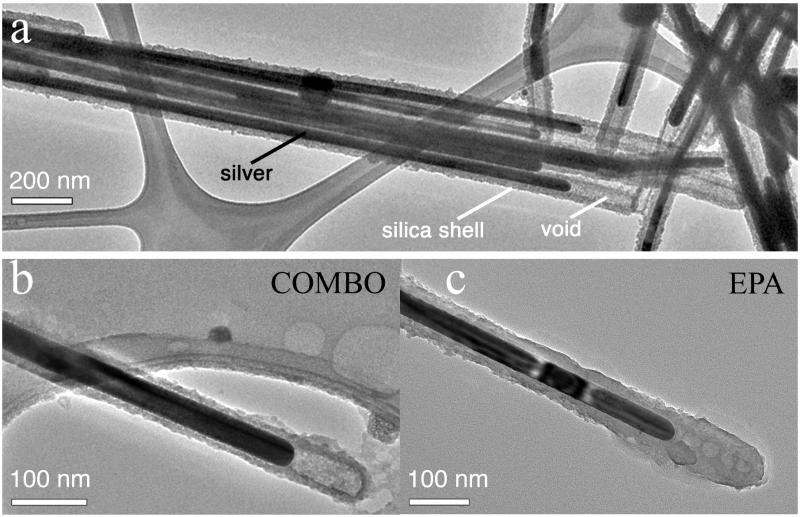
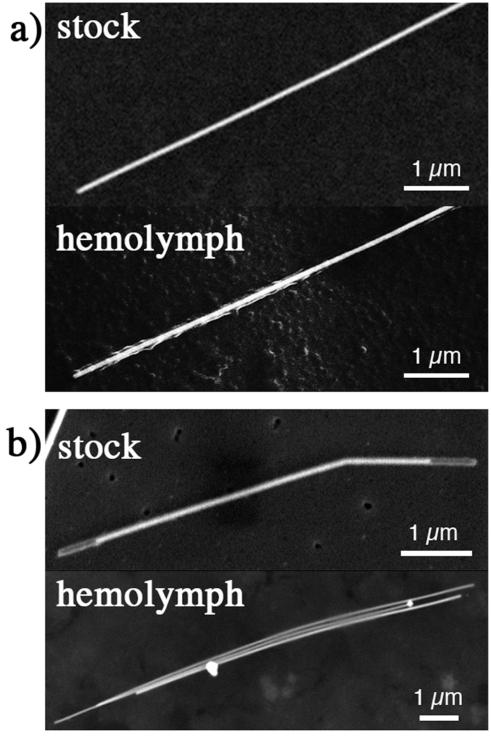
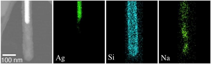
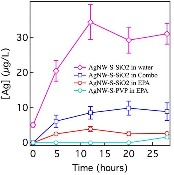
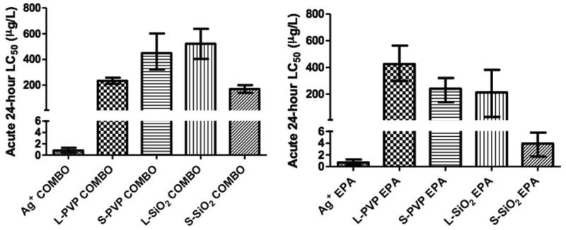
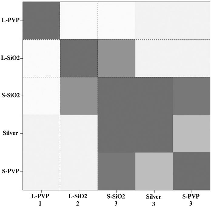
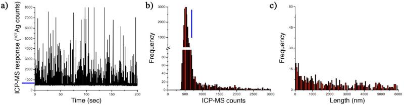
Nanowires (NWs), high-aspect-ratio nanomaterials, are increasingly used in technological materials and consumer products and may have toxicological characteristics distinct from nanoparticles. We carried out a comprehensive evaluation of the physicochemical stability of four silver nanowires (AgNWs) of two sizes and coatings and their toxicity to Daphnia magna . Inorganic aluminum-doped silica coatings were less effective than organic poly(vinyl pyrrolidone) coatings at preventing silver oxidation or Ag(+) release and underwent a significant morphological transformation within 1 h following addition to low ionic strength Daphnia growth media. All AgNWs were highly toxic to D. magna but less toxic than ionic silver. Toxicity varied as a function of AgNW dimension, coating, and solution chemistry. Ag(+) release in the media could not account for observed AgNW toxicity. Single-particle inductively coupled plasma mass spectrometry distinguished and quantified dissolved and nanoparticulate silver in microliter-scale volumes of Daphnia magna hemolymph with a limit of detection of approximately 10 ppb. The silver levels within the hemolymph of Daphnia exposed to both Ag(+) and AgNW met or exceeded the initial concentration in the growth medium, indicating effective accumulation during filter feeding. Silver-rich particles were the predominant form of silver in hemolymph following exposure to both AgNWs and Ag(+). Scanning electron microscopy imaging of dried hemolymph found both AgNWs and silver precipitates that were not present in the AgNW stock or the growth medium. Both organic and inorganic coatings on the AgNW were transformed during ingestion or absorption. Pathway, gene ontology, and clustering analyses of gene expression response indicated effects of AgNWs distinct from ionic silver on Daphnia magna .

摘要

纳米线(NWs)作为高纵横比的纳米材料,在技术材料和消费品中的应用日益广泛,其毒理学特性可能与纳米颗粒不同。我们对两种尺寸和涂层的四种银纳米线(AgNWs)的物理化学稳定性及其对大型溞的毒性进行了全面评估。在防止银氧化或银离子(Ag(+))释放方面,无机铝掺杂二氧化硅涂层的效果不如有机聚乙烯吡咯烷酮涂层,并且在添加到低离子强度的大型溞生长培养基中1小时内会发生显著的形态转变。所有AgNWs对大型溞都具有高毒性,但毒性低于离子银。毒性随AgNW尺寸、涂层和溶液化学性质而变化。培养基中的Ag(+)释放不能解释观察到的AgNW毒性。单颗粒电感耦合等离子体质谱法能够区分和定量微升规模的大型溞血淋巴中的溶解态和纳米颗粒态银,检测限约为10 ppb。暴露于Ag(+)和AgNW的大型溞血淋巴中的银含量达到或超过了生长培养基中的初始浓度,表明在滤食过程中有效积累。暴露于AgNWs和Ag(+)后,富含银的颗粒是血淋巴中银的主要形式。对干燥血淋巴的扫描电子显微镜成像发现了AgNWs和银沉淀物,这些在AgNW储备液或生长培养基中并不存在。AgNW上的有机和无机涂层在摄入或吸收过程中都会发生转变。基因表达反应的通路、基因本体和聚类分析表明,AgNWs对大型溞的影响与离子银不同。

https://cdn.ncbi.nlm.nih.gov/pmc/blobs/5817/3912856/e1e13f2d6e15/nihms-547078-f0007.jpg
https://cdn.ncbi.nlm.nih.gov/pmc/blobs/5817/3912856/4cbd9685906f/nihms-547078-f0001.jpg
https://cdn.ncbi.nlm.nih.gov/pmc/blobs/5817/3912856/4b6261b90597/nihms-547078-f0002.jpg
https://cdn.ncbi.nlm.nih.gov/pmc/blobs/5817/3912856/85423508bfec/nihms-547078-f0003.jpg
https://cdn.ncbi.nlm.nih.gov/pmc/blobs/5817/3912856/3526bf665223/nihms-547078-f0004.jpg
https://cdn.ncbi.nlm.nih.gov/pmc/blobs/5817/3912856/4d509fb3bf59/nihms-547078-f0005.jpg
https://cdn.ncbi.nlm.nih.gov/pmc/blobs/5817/3912856/3ceab476b202/nihms-547078-f0006.jpg
https://cdn.ncbi.nlm.nih.gov/pmc/blobs/5817/3912856/e1e13f2d6e15/nihms-547078-f0007.jpg