• 文献检索
  • 文档翻译
  • 深度研究
  • 学术资讯
  • Suppr Zotero 插件Zotero 插件
  • 邀请有礼
  • 套餐&价格
  • 历史记录
应用&插件
Suppr Zotero 插件Zotero 插件浏览器插件Mac 客户端Windows 客户端微信小程序
定价
高级版会员购买积分包购买API积分包
服务
文献检索文档翻译深度研究API 文档MCP 服务
关于我们
关于 Suppr公司介绍联系我们用户协议隐私条款
关注我们

Suppr 超能文献

核心技术专利:CN118964589B侵权必究
粤ICP备2023148730 号-1Suppr @ 2026

文献检索

告别复杂PubMed语法,用中文像聊天一样搜索,搜遍4000万医学文献。AI智能推荐,让科研检索更轻松。

立即免费搜索

文件翻译

保留排版,准确专业,支持PDF/Word/PPT等文件格式,支持 12+语言互译。

免费翻译文档

深度研究

AI帮你快速写综述,25分钟生成高质量综述,智能提取关键信息,辅助科研写作。

立即免费体验

耳鸣患者的听觉网络连通性:一项静息态功能磁共振成像研究。

Auditory network connectivity in tinnitus patients: a resting-state fMRI study.

作者信息

Davies J, Gander P E, Andrews M, Hall D A

机构信息

* NIHR Nottingham Hearing Biomedical Research Unit, University of Nottingham , UK.

出版信息

Int J Audiol. 2014 Mar;53(3):192-8. doi: 10.3109/14992027.2013.846482. Epub 2013 Nov 7.

DOI:10.3109/14992027.2013.846482
PMID:24200464
原文链接:https://pmc.ncbi.nlm.nih.gov/articles/PMC3956492/
Abstract

OBJECTIVE

Resting-state functional magnetic resonance imaging (fMRI) uncovers correlated activity between spatially distinct functionally related brain regions and offers clues about the integrity of functional brain circuits in people with chronic subjective tinnitus. We chose to investigate auditory network connectivity, adopting and extending previously used analyses methods to provide an independent evaluation of replicability.

DESIGN

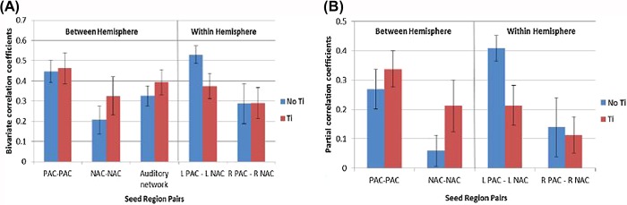
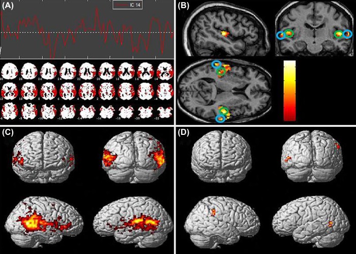
Independent components analysis (ICA) was used to identify coherent patterns arising from spontaneous brain signals within the resting-state data. The auditory network component was extracted and evaluated. Bivariate and partial correlation analyses were performed on pre-defined regions of bilateral auditory cortex to assess functional connectivity.

STUDY SAMPLE

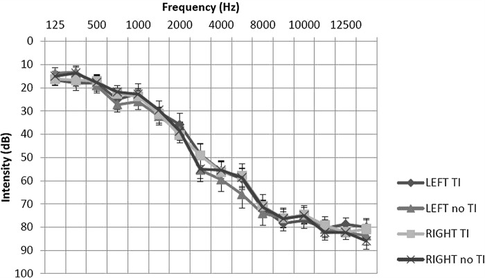
Our design carefully matched participant groups for possible confounds, such as hearing status. Twelve patients (seven male, five female; mean age 66 years) all with chronic constant tinnitus and eleven controls (eight male, three female; mean age 68 years) took part.

RESULTS

No significant differences were found in auditory network connectivity between groups after correcting for multiple statistical comparisons in the analysis. This contradicts previous findings reporting reduced auditory network connectivity; albeit at a less stringent statistical threshold.

CONCLUSIONS

Auditory network connectivity does not appear to be reliably altered by the experience of chronic subjective tinnitus.

摘要

目的

静息态功能磁共振成像(fMRI)揭示了空间上不同的功能相关脑区之间的相关活动,并为慢性主观性耳鸣患者功能性脑回路的完整性提供了线索。我们选择研究听觉网络连通性,采用并扩展先前使用的分析方法以对可重复性进行独立评估。

设计

独立成分分析(ICA)用于识别静息态数据中自发脑信号产生的连贯模式。提取并评估听觉网络成分。对双侧听觉皮层的预定义区域进行双变量和偏相关分析,以评估功能连通性。

研究样本

我们的设计仔细匹配了参与者组,以排除可能的混杂因素,如听力状况。12名患者(7名男性,5名女性;平均年龄66岁)均患有慢性持续性耳鸣,11名对照者(8名男性,3名女性;平均年龄68岁)参与了研究。

结果

在分析中进行多重统计比较校正后,两组之间的听觉网络连通性未发现显著差异。这与先前报道听觉网络连通性降低的研究结果相矛盾;尽管先前研究的统计阈值没那么严格。

结论

慢性主观性耳鸣似乎并未可靠地改变听觉网络连通性。

https://cdn.ncbi.nlm.nih.gov/pmc/blobs/61fa/3956492/a43ed4fd0956/IJA-53-192-g003.jpg
https://cdn.ncbi.nlm.nih.gov/pmc/blobs/61fa/3956492/aa4a7bc6e120/IJA-53-192-g001.jpg
https://cdn.ncbi.nlm.nih.gov/pmc/blobs/61fa/3956492/4296c3037729/IJA-53-192-g002.jpg
https://cdn.ncbi.nlm.nih.gov/pmc/blobs/61fa/3956492/a43ed4fd0956/IJA-53-192-g003.jpg
https://cdn.ncbi.nlm.nih.gov/pmc/blobs/61fa/3956492/aa4a7bc6e120/IJA-53-192-g001.jpg
https://cdn.ncbi.nlm.nih.gov/pmc/blobs/61fa/3956492/4296c3037729/IJA-53-192-g002.jpg
https://cdn.ncbi.nlm.nih.gov/pmc/blobs/61fa/3956492/a43ed4fd0956/IJA-53-192-g003.jpg

相似文献

1
Auditory network connectivity in tinnitus patients: a resting-state fMRI study.耳鸣患者的听觉网络连通性:一项静息态功能磁共振成像研究。
Int J Audiol. 2014 Mar;53(3):192-8. doi: 10.3109/14992027.2013.846482. Epub 2013 Nov 7.
2
Using resting state functional connectivity to unravel networks of tinnitus.利用静息态功能连接来揭示耳鸣的网络。
Hear Res. 2014 Jan;307:153-62. doi: 10.1016/j.heares.2013.07.010. Epub 2013 Jul 26.
3
Connectivity graph analysis of the auditory resting state network in tinnitus.耳鸣静息态网络的连通性图谱分析。
Brain Res. 2012 Nov 16;1485:10-21. doi: 10.1016/j.brainres.2012.05.006. Epub 2012 May 10.
4
Alteration of functional connectivity in tinnitus brain revealed by resting-state fMRI? A pilot study.静息态 fMRI 显示耳鸣脑功能连接的改变?一项初步研究。
Int J Audiol. 2012 May;51(5):413-7. doi: 10.3109/14992027.2011.652677. Epub 2012 Jan 30.
5
Default mode, dorsal attention and auditory resting state networks exhibit differential functional connectivity in tinnitus and hearing loss.静息态默认模式网络、背侧注意网络和听觉网络在耳鸣和听力损失中的功能连接存在差异。
PLoS One. 2013 Oct 2;8(10):e76488. doi: 10.1371/journal.pone.0076488. eCollection 2013.
6
Tinnitus distress is linked to enhanced resting-state functional connectivity from the limbic system to the auditory cortex.耳鸣困扰与从边缘系统到听觉皮层的静息态功能连接增强有关。
Hum Brain Mapp. 2017 May;38(5):2384-2397. doi: 10.1002/hbm.23525. Epub 2017 Jan 23.
7
A 7 Tesla fMRI investigation of human tinnitus percept in cortical and subcortical auditory areas.7 特斯拉 fMRI 研究人类耳鸣感知在皮质和皮质下听觉区域的表现。
Neuroimage Clin. 2020;25:102166. doi: 10.1016/j.nicl.2020.102166. Epub 2020 Jan 11.
8
Auditory resting-state functional connectivity in tinnitus and modulation with transcranial direct current stimulation.耳鸣中的听觉静息态功能连接及经颅直流电刺激的调节作用
Acta Otolaryngol. 2015;135(12):1286-92. doi: 10.3109/00016489.2015.1068952. Epub 2015 Jul 16.
9
Reduced sound-evoked and resting-state BOLD fMRI connectivity in tinnitus.耳鸣患者听觉诱发和静息状态 fMRI 连接的减少。
Neuroimage Clin. 2018 Aug 31;20:637-649. doi: 10.1016/j.nicl.2018.08.029. eCollection 2018.
10
Dynamic Changes of Functional Neuronal Activities Between the Auditory Pathway and Limbic Systems Contribute to Noise-Induced Tinnitus with a Normal Audiogram.听觉通路和边缘系统之间功能性神经元活动的动态变化导致伴有正常听阈的噪声性耳鸣。
Neuroscience. 2019 Jun 1;408:31-45. doi: 10.1016/j.neuroscience.2019.03.054. Epub 2019 Apr 1.

引用本文的文献

1
An examination of the reliability of seed-to-seed resting state functional connectivity in tinnitus patients.耳鸣患者种子到种子静息态功能连接的可靠性研究。
Neuroimage Rep. 2023 Feb 15;3(1):100158. doi: 10.1016/j.ynirp.2023.100158. eCollection 2023 Mar.
2
[Progress in neural network mechanism of tinnitus using functional magnetic resonance imaging].[利用功能磁共振成像技术对耳鸣神经网络机制的研究进展]
Lin Chuang Er Bi Yan Hou Tou Jing Wai Ke Za Zhi. 2023 Jul;37(7):582-587. doi: 10.13201/j.issn.2096-7993.2023.07.014.
3
Intrinsic Network Changes in Bilateral Tinnitus Patients with Cognitive Impairment: A Resting-State Functional MRI Study.

本文引用的文献

1
Tinnitus.耳鸣。
Lancet. 2013 Nov 9;382(9904):1600-7. doi: 10.1016/S0140-6736(13)60142-7. Epub 2013 Jul 2.
2
Methodological aspects of clinical trials in tinnitus: a proposal for an international standard.耳鸣临床试验的方法学方面:国际标准建议。
J Psychosom Res. 2012 Aug;73(2):112-21. doi: 10.1016/j.jpsychores.2012.05.002. Epub 2012 Jun 22.
3
Functional connectivity networks in nonbothersome tinnitus.无扰性耳鸣的功能连接网络。
伴有认知障碍的双侧耳鸣患者的内在网络变化:一项静息态功能磁共振成像研究
Brain Sci. 2022 Aug 8;12(8):1049. doi: 10.3390/brainsci12081049.
4
Disrupted intra- and inter-network connectivity in unilateral acute tinnitus with hearing loss.单侧急性耳鸣伴听力损失时,脑内及脑间网络连接中断。
Front Aging Neurosci. 2022 Aug 1;14:833437. doi: 10.3389/fnagi.2022.833437. eCollection 2022.
5
Resting-state Networks in Tinnitus : A Scoping Review.耳鸣的静息态网络:一项范围综述。
Clin Neuroradiol. 2022 Dec;32(4):903-922. doi: 10.1007/s00062-022-01170-1. Epub 2022 May 12.
6
The Neural Mechanisms of Tinnitus: A Perspective From Functional Magnetic Resonance Imaging.耳鸣的神经机制:来自功能磁共振成像的视角
Front Neurosci. 2021 Feb 11;15:621145. doi: 10.3389/fnins.2021.621145. eCollection 2021.
7
Comparing Cyclicity Analysis With Pre-established Functional Connectivity Methods to Identify Individuals and Subject Groups Using Resting State fMRI.将周期性分析与预先建立的功能连接方法进行比较,以使用静息态功能磁共振成像识别个体和受试者组。
Front Comput Neurosci. 2020 Jan 20;13:94. doi: 10.3389/fncom.2019.00094. eCollection 2019.
8
Abnormal Spontaneous Neural Activity of the Central Auditory System Changes the Functional Connectivity in the Tinnitus Brain: A Resting-State Functional MRI Study.中枢听觉系统异常的自发神经活动改变耳鸣大脑中的功能连接:一项静息态功能磁共振成像研究
Front Neurosci. 2019 Dec 20;13:1314. doi: 10.3389/fnins.2019.01314. eCollection 2019.
9
Maladaptive alterations of resting state cortical network in Tinnitus: A directed functional connectivity analysis of a larger MEG data set.耳鸣静息态皮质网络的适应不良改变:更大的 MEG 数据集的有向功能连接分析。
Sci Rep. 2019 Oct 29;9(1):15452. doi: 10.1038/s41598-019-51747-z.
10
Functional Near-Infrared Spectroscopy to Probe tDCS-Induced Cortical Functioning Changes in Tinnitus.利用功能近红外光谱技术探究经颅直流电刺激诱发的耳鸣患者皮质功能变化
J Int Adv Otol. 2019 Aug;15(2):321-325. doi: 10.5152/iao.2019.6022.
Otolaryngol Head Neck Surg. 2012 Nov;147(5):900-6. doi: 10.1177/0194599812451414. Epub 2012 Jun 21.
4
Functional connectivity during modulation of tinnitus with orofacial maneuvers.经口运动调节耳鸣时的功能连接。
Otolaryngol Head Neck Surg. 2012 Oct;147(4):757-62. doi: 10.1177/0194599812450680. Epub 2012 Jun 6.
5
Connectivity graph analysis of the auditory resting state network in tinnitus.耳鸣静息态网络的连通性图谱分析。
Brain Res. 2012 Nov 16;1485:10-21. doi: 10.1016/j.brainres.2012.05.006. Epub 2012 May 10.
6
Auditory resting-state network connectivity in tinnitus: a functional MRI study.耳鸣的听觉静息态网络连接:一项功能磁共振成像研究。
PLoS One. 2012;7(5):e36222. doi: 10.1371/journal.pone.0036222. Epub 2012 May 4.
7
Subcallosal brain structure: correlation with hearing threshold at supra-clinical frequencies (>8 kHz), but not with tinnitus.胼胝体下回脑结构:与超临床频率(>8 kHz)的听力阈值相关,而与耳鸣无关。
Hear Res. 2013 Jan;295:79-86. doi: 10.1016/j.heares.2012.03.013. Epub 2012 Apr 7.
8
Neural correlates of tinnitus related distress: an fMRI-study.耳鸣相关困扰的神经相关因素:一项 fMRI 研究。
Hear Res. 2013 Jan;295:87-99. doi: 10.1016/j.heares.2012.03.003. Epub 2012 Mar 14.
9
Alteration of functional connectivity in tinnitus brain revealed by resting-state fMRI? A pilot study.静息态 fMRI 显示耳鸣脑功能连接的改变?一项初步研究。
Int J Audiol. 2012 May;51(5):413-7. doi: 10.3109/14992027.2011.652677. Epub 2012 Jan 30.
10
Altered networks in bothersome tinnitus: a functional connectivity study.恼人耳鸣的改变网络:一项功能连接研究。
BMC Neurosci. 2012 Jan 4;13:3. doi: 10.1186/1471-2202-13-3.