Key Laboratory of the Ministry of Education for Experimental Teratology, Shandong Provincial Key Laboratory of Mental Disorders, Department of Histology and Embryology, Shandong University School of Medicine, Jinan, Shandong, China ; Department of Anatomy, Yong Loo Lin School of Medicine, National University of Singapore, Singapore, Singapore.
PLoS One. 2013 Nov 6;8(11):e78439. doi: 10.1371/journal.pone.0078439. eCollection 2013.
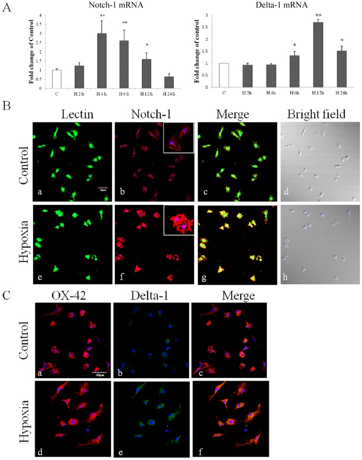
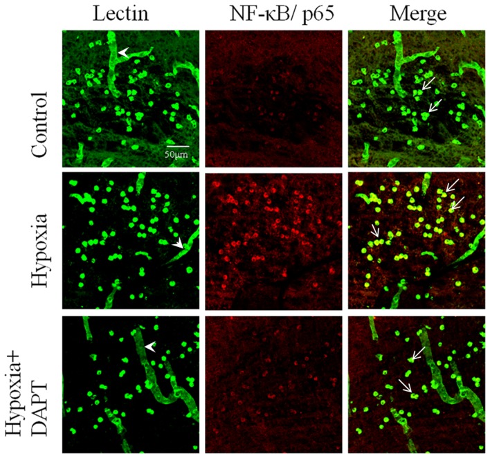
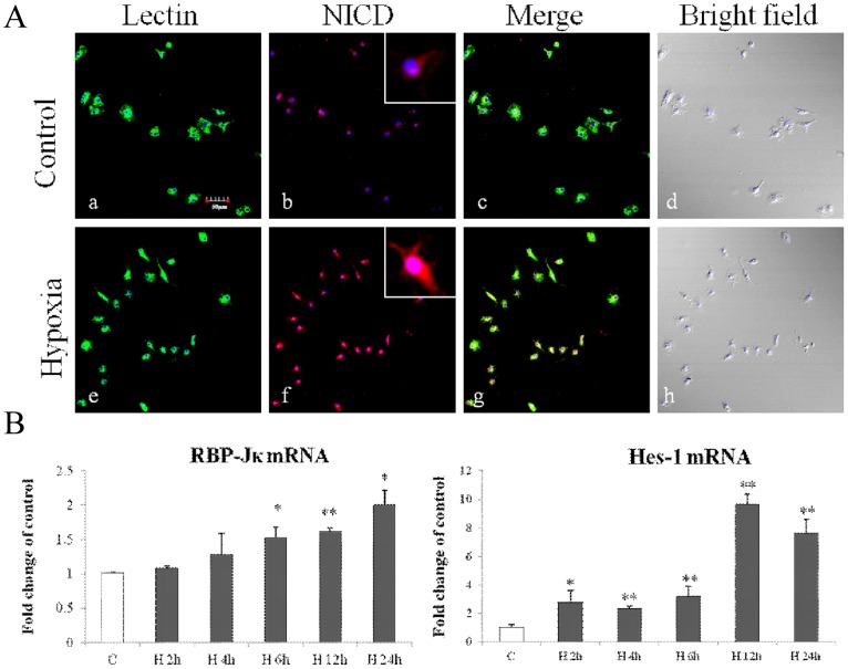
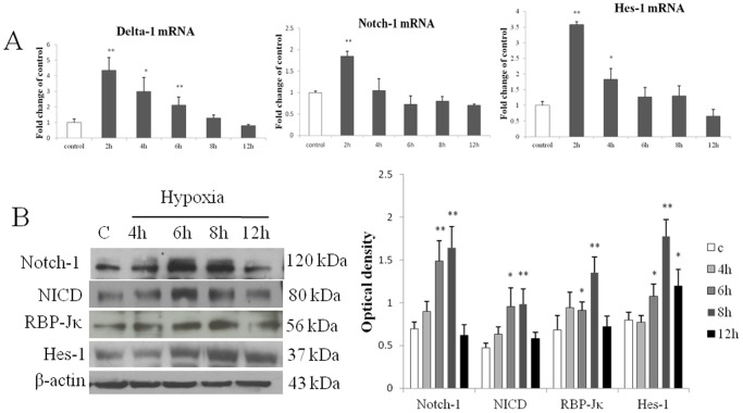
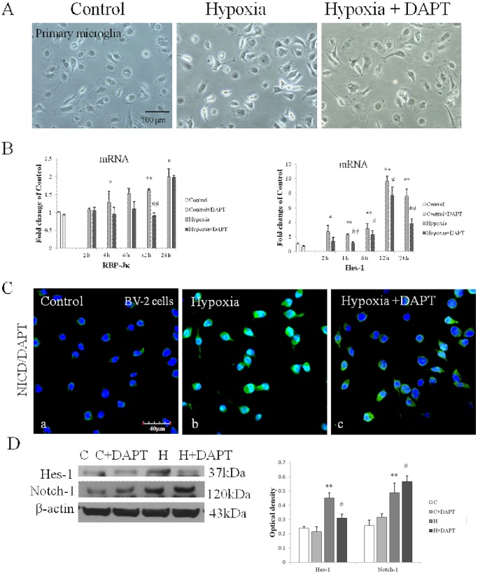
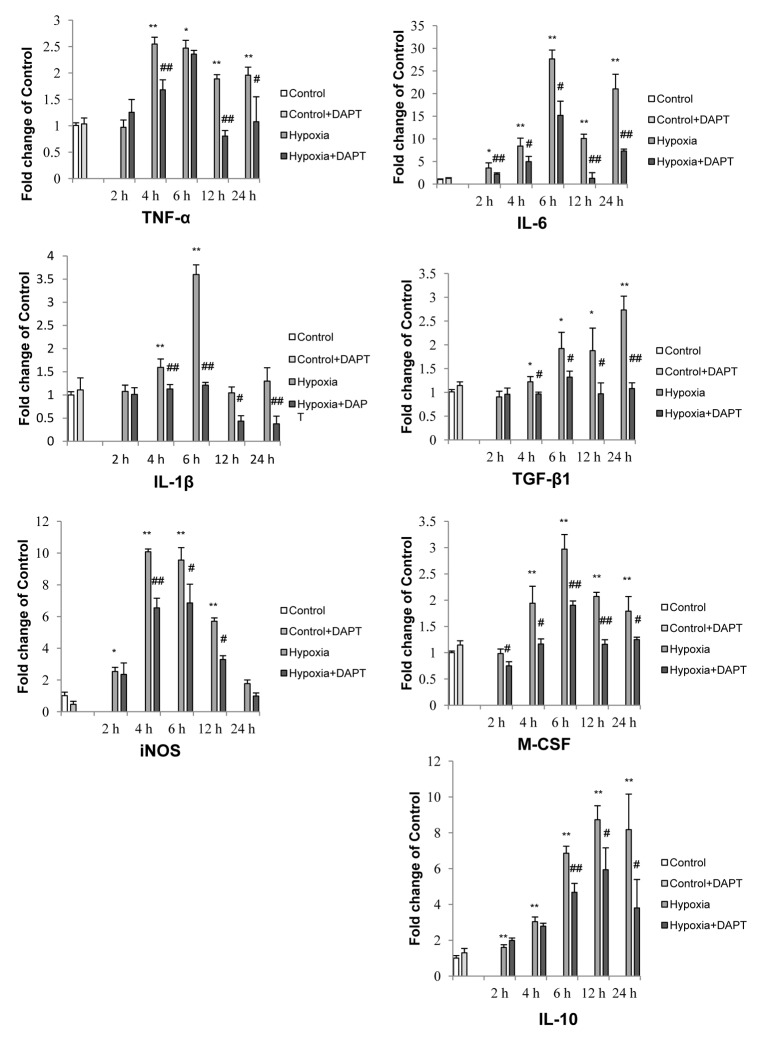
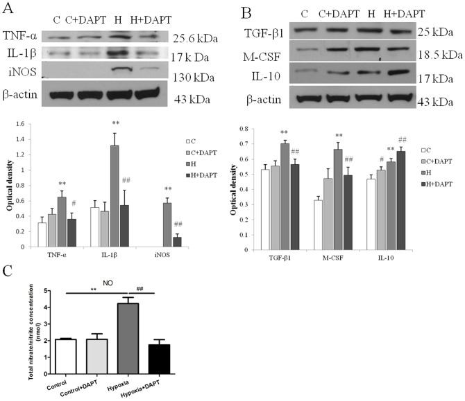
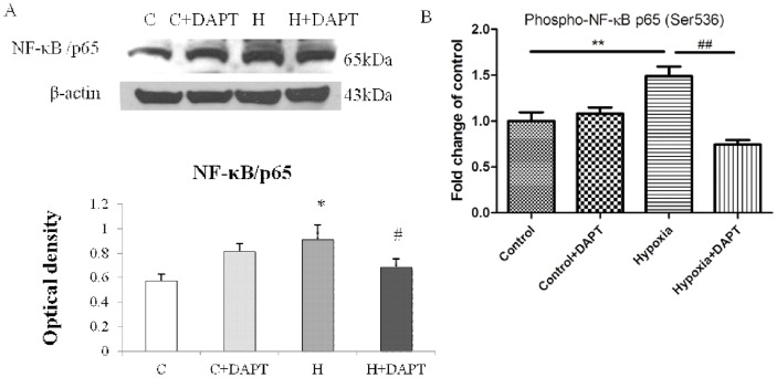
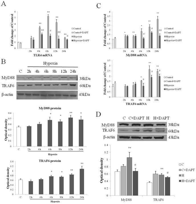
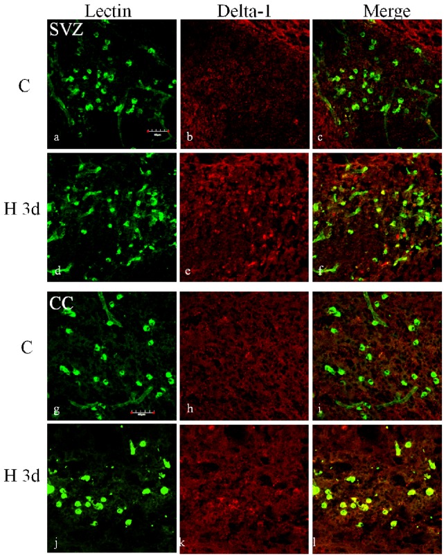
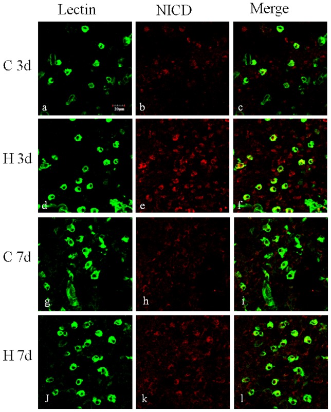
Neuroinflammation mediated by the activated microglia is suggested to play a pivotal role in the pathogenesis of hypoxic brain injury; however, the underlying mechanism of microglia activation remains unclear. Here, we show that the canonical Notch signaling orchestrates microglia activation after hypoxic exposure which is closely associated with multiple pathological situations of the brain. Notch-1 and Delta-1 expression in primary microglia and BV-2 microglial cells was significantly elevated after hypoxia. Hypoxia-induced activation of Notch signaling was further confirmed by the concomitant increase in the expression and translocation of intracellular Notch receptor domain (NICD), together with RBP-Jκ and target gene Hes-1 expression. Chemical inhibition of Notch signaling with N-[N-(3,5-difluorophenacetyl)-1-alany1- S-phenyglycine t-butyl ester (DAPT), a γ-secretase inhibitor, effectively reduced hypoxia-induced upregulated expression of most inflammatory mediators. Notch inhibition also reduced NF-κB/p65 expression and translocation. Remarkably, Notch inhibition suppressed expression of TLR4/MyD88/TRAF6 pathways. In vivo, Notch signaling expression and activation in microglia were observed in the cerebrum of postnatal rats after hypoxic injury. Most interestingly, hypoxia-induced upregulation of NF-κB immunoexpression in microglia was prevented when the rats were given DAPT pretreatment underscoring the interrelationship between Notch signaling and NF-κB pathways. Taken together, we conclude that Notch signaling is involved in regulating microglia activation after hypoxia partly through the cross talk between TLR4/MyD88/TRAF6/NF-κB pathways. Therefore, Notch signaling may serve as a prospective target for inhibition of microglia activation known to be implicated in brain damage in the developing brain.
被激活的小胶质细胞介导的神经炎症被认为在缺氧性脑损伤发病机制中起关键作用;然而,小胶质细胞激活的潜在机制尚不清楚。在这里,我们表明经典的 Notch 信号转导在缺氧暴露后协调小胶质细胞的激活,这与大脑的多种病理情况密切相关。初级小胶质细胞和 BV-2 小胶质细胞中 Notch-1 和 Delta-1 的表达在缺氧后显著升高。 Notch 信号转导的缺氧诱导激活进一步通过细胞内 Notch 受体结构域(NICD)的表达和易位的伴随增加来证实,同时伴随着 RBP-Jκ 和靶基因 Hes-1 的表达。 Notch 信号通路的化学抑制,用 N-[N-(3,5-二氟苯乙酰基)-1-丙氨酸-S-苯甘氨酸叔丁酯(DAPT),一种γ-分泌酶抑制剂,有效地降低了缺氧诱导的大多数炎症介质的上调表达。 Notch 抑制也降低了 NF-κB/p65 的表达和易位。值得注意的是, Notch 抑制抑制了 TLR4/MyD88/TRAF6 通路的表达。在体内,在缺氧损伤后新生大鼠大脑中的小胶质细胞中观察到 Notch 信号表达和激活。最有趣的是,当大鼠给予 DAPT 预处理时,缺氧诱导的小胶质细胞中 NF-κB 免疫表达的上调被阻止,这强调了 Notch 信号和 NF-κB 通路之间的相互关系。总之,我们得出结论, Notch 信号转导参与调节缺氧后小胶质细胞的激活,部分通过 TLR4/MyD88/TRAF6/NF-κB 通路的串扰。因此, Notch 信号转导可能作为一个有前途的抑制小胶质细胞激活的靶点,已知小胶质细胞激活与发育中的大脑损伤有关。