• 文献检索
  • 文档翻译
  • 深度研究
  • 学术资讯
  • Suppr Zotero 插件Zotero 插件
  • 邀请有礼
  • 套餐&价格
  • 历史记录
应用&插件
Suppr Zotero 插件Zotero 插件浏览器插件Mac 客户端Windows 客户端微信小程序
定价
高级版会员购买积分包购买API积分包
服务
文献检索文档翻译深度研究API 文档MCP 服务
关于我们
关于 Suppr公司介绍联系我们用户协议隐私条款
关注我们

Suppr 超能文献

核心技术专利:CN118964589B侵权必究
粤ICP备2023148730 号-1Suppr @ 2026

文献检索

告别复杂PubMed语法,用中文像聊天一样搜索,搜遍4000万医学文献。AI智能推荐,让科研检索更轻松。

立即免费搜索

文件翻译

保留排版,准确专业,支持PDF/Word/PPT等文件格式,支持 12+语言互译。

免费翻译文档

深度研究

AI帮你快速写综述,25分钟生成高质量综述,智能提取关键信息,辅助科研写作。

立即免费体验

乳腺MRI中小病变的分类:通过特征选择评估动态提取纹理特征的作用。

Classification of Small Lesions in Breast MRI: Evaluating The Role of Dynamically Extracted Texture Features Through Feature Selection.

作者信息

Nagarajan Mahesh B, Huber Markus B, Schlossbauer Thomas, Leinsinger Gerda, Krol Andrzej, Wismüller Axel

机构信息

Departments of Imaging Sciences and Biomedical Engineering, University of Rochester, Rochester NY 14627, USA.

出版信息

J Med Biol Eng. 2013 Jan 1;33(1). doi: 10.5405/jmbe.1183.

DOI:10.5405/jmbe.1183
PMID:24223533
原文链接:https://pmc.ncbi.nlm.nih.gov/articles/PMC3820163/
Abstract

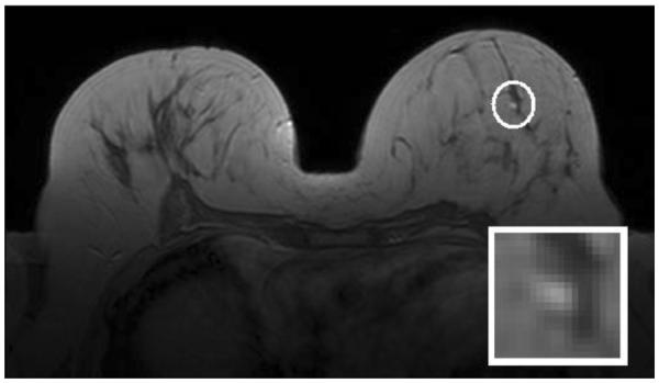
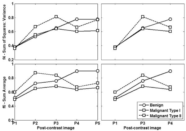
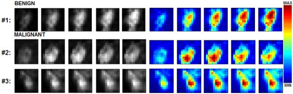
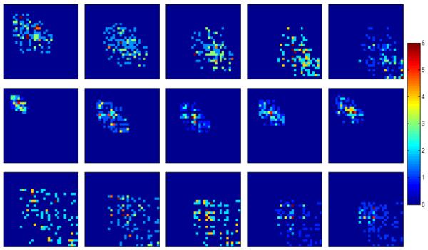
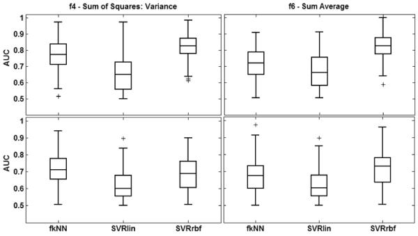
Dynamic texture quantification, i.e., extracting texture features from the lesion enhancement pattern in all available post-contrast images, has not been evaluated in terms of its ability to classify small lesions. This study investigates the classification performance achieved with texture features extracted from all five post-contrast images of lesions (mean lesion diameter of 1.1 cm) annotated in dynamic breast magnetic resonance imaging exams. Sixty lesions are characterized dynamically using Haralick texture features. The texture features are then used in a classification task with support vector regression and a fuzzy k-nearest neighbor classifier; free parameters of these classifiers are optimized using random sub-sampling cross-validation. Classifier performance is determined through receiver-operator characteristic (ROC) analysis, specifically through computation of the area under the ROC curve (AUC). Mutual information is used to evaluate the contribution of texture features extracted from different post-contrast stages to classifier performance. Significant improvements (p < 0.05) are observed for six of the thirteen texture features when the lesion enhancement pattern is quantified using the proposed approach of dynamic texture quantification. The highest AUC value observed (0.82) is achieved with texture features responsible for capturing aspects of lesion heterogeneity. Mutual information analysis reveals that texture features extracted from the third and fourth post-contrast images contributed most to the observed improvement in classifier performance. These results show that the performance of automated character classification with small lesions can be significantly improved through dynamic texture quantification of the lesion enhancement pattern.

摘要

动态纹理量化,即从所有可用的增强后图像中的病变增强模式中提取纹理特征,尚未就其对小病变进行分类的能力进行评估。本研究调查了从动态乳腺磁共振成像检查中标注的病变(平均病变直径为1.1厘米)的所有五幅增强后图像中提取的纹理特征所实现的分类性能。使用哈氏纹理特征对60个病变进行动态特征描述。然后将纹理特征用于支持向量回归和模糊k近邻分类器的分类任务;使用随机子采样交叉验证对这些分类器的自由参数进行优化。通过接收者操作特征(ROC)分析,特别是通过计算ROC曲线下面积(AUC)来确定分类器性能。互信息用于评估从不同增强后阶段提取的纹理特征对分类器性能的贡献。当使用所提出的动态纹理量化方法对病变增强模式进行量化时,13个纹理特征中的6个有显著改善(p < 0.05)。观察到的最高AUC值(0.82)是通过负责捕捉病变异质性方面的纹理特征实现的。互信息分析表明,从第三和第四幅增强后图像中提取的纹理特征对观察到的分类器性能改善贡献最大。这些结果表明,通过对病变增强模式进行动态纹理量化,可以显著提高小病变自动特征分类的性能。

https://cdn.ncbi.nlm.nih.gov/pmc/blobs/d8a3/3820163/506005d72a14/nihms-482680-f0006.jpg
https://cdn.ncbi.nlm.nih.gov/pmc/blobs/d8a3/3820163/d88f0190ddc3/nihms-482680-f0001.jpg
https://cdn.ncbi.nlm.nih.gov/pmc/blobs/d8a3/3820163/5f8b172333a0/nihms-482680-f0002.jpg
https://cdn.ncbi.nlm.nih.gov/pmc/blobs/d8a3/3820163/833252267481/nihms-482680-f0003.jpg
https://cdn.ncbi.nlm.nih.gov/pmc/blobs/d8a3/3820163/61656672c920/nihms-482680-f0004.jpg
https://cdn.ncbi.nlm.nih.gov/pmc/blobs/d8a3/3820163/35954e23af60/nihms-482680-f0005.jpg
https://cdn.ncbi.nlm.nih.gov/pmc/blobs/d8a3/3820163/506005d72a14/nihms-482680-f0006.jpg

相似文献

1
Classification of Small Lesions in Breast MRI: Evaluating The Role of Dynamically Extracted Texture Features Through Feature Selection.乳腺MRI中小病变的分类:通过特征选择评估动态提取纹理特征的作用。
J Med Biol Eng. 2013 Jan 1;33(1). doi: 10.5405/jmbe.1183.
2
Classification of small lesions in dynamic breast MRI: Eliminating the need for precise lesion segmentation through spatio-temporal analysis of contrast enhancement over time.动态乳腺磁共振成像中小病变的分类:通过对随时间变化的对比增强进行时空分析,无需精确的病变分割。
Mach Vis Appl. 2013 Oct 1;24(7). doi: 10.1007/s00138-012-0456-y.
3
Multi-parametric MRI lesion heterogeneity biomarkers for breast cancer diagnosis.多参数 MRI 病变异质性生物标志物在乳腺癌诊断中的应用。
Phys Med. 2020 Dec;80:101-110. doi: 10.1016/j.ejmp.2020.10.007. Epub 2020 Nov 1.
4
Classification of small lesions on dynamic breast MRI: Integrating dimension reduction and out-of-sample extension into CADx methodology.动态乳腺 MRI 中小病灶的分类:将降维和样本外扩展纳入 CADx 方法。
Artif Intell Med. 2014 Jan;60(1):65-77. doi: 10.1016/j.artmed.2013.11.003. Epub 2013 Nov 23.
5
Differentiation of fat-poor angiomyolipoma from clear cell renal cell carcinoma in contrast-enhanced MDCT images using quantitative feature classification.基于定量特征分类的 MDCT 增强图像鉴别乏脂性血管平滑肌脂肪瘤与透明细胞肾细胞癌
Med Phys. 2017 Jul;44(7):3604-3614. doi: 10.1002/mp.12258. Epub 2017 Jun 9.
6
Quantitative analysis of lesion morphology and texture features for diagnostic prediction in breast MRI.乳腺MRI中病变形态和纹理特征的定量分析用于诊断预测
Acad Radiol. 2008 Dec;15(12):1513-25. doi: 10.1016/j.acra.2008.06.005.
7
Investigating the use of texture features for analysis of breast lesions on contrast-enhanced cone beam CT.研究纹理特征在对比增强锥形束CT上分析乳腺病变中的应用。
Proc SPIE Int Soc Opt Eng. 2014 Apr 9;9038. doi: 10.1117/12.2042397.
8
Assessing heterogeneity of lesion enhancement kinetics in dynamic contrast-enhanced MRI for breast cancer diagnosis.评估动态对比增强 MRI 中病变增强动力学的异质性用于乳腺癌诊断。
Br J Radiol. 2010 Apr;83(988):296-309. doi: 10.1259/bjr/50743919.
9
Textural analysis of early-phase spatiotemporal changes in contrast enhancement of breast lesions imaged with an ultrafast DCE-MRI protocol.利用超快 DCE-MRI 协议对乳腺病变的早期时空调制对比增强进行纹理分析。
Med Phys. 2017 Sep;44(9):4652-4664. doi: 10.1002/mp.12408. Epub 2017 Jul 18.
10
Characterization of spatiotemporal changes for the classification of dynamic contrast-enhanced magnetic-resonance breast lesions.动态对比增强磁共振乳腺病变分类的时空变化特征。
Artif Intell Med. 2013 Jun;58(2):101-14. doi: 10.1016/j.artmed.2013.03.002. Epub 2013 Mar 30.

引用本文的文献

1
Vascularity and Dynamic Contrast-Enhanced Breast Magnetic Resonance Imaging.血管造影与动态对比增强乳腺磁共振成像
Front Radiol. 2021 Dec 9;1:735567. doi: 10.3389/fradi.2021.735567. eCollection 2021.
2
A Pipelined Tracer-Aware Approach for Lesion Segmentation in Breast DCE-MRI.一种用于乳腺动态对比增强磁共振成像中病变分割的流水线式跟踪器感知方法。
J Imaging. 2021 Dec 14;7(12):276. doi: 10.3390/jimaging7120276.
3
Association between breast cancer's prognostic factors and 3D textural features of non-contrast-enhanced weighted breast MRI.

本文引用的文献

1
Textural kinetics: a novel dynamic contrast-enhanced (DCE)-MRI feature for breast lesion classification.纹理动力学:一种新的动态对比增强(DCE)MRI 特征,用于乳腺病变分类。
J Digit Imaging. 2011 Jun;24(3):446-63. doi: 10.1007/s10278-010-9298-1.
2
STEP: spatiotemporal enhancement pattern for MR-based breast tumor diagnosis.步骤:基于磁共振成像的乳腺肿瘤诊断的时空增强模式
Med Phys. 2009 Jul;36(7):3192-204. doi: 10.1118/1.3151811.
3
Quantitative analysis of lesion morphology and texture features for diagnostic prediction in breast MRI.
乳腺癌预后因素与非增强加权乳腺 MRI 的 3D 纹理特征之间的关系。
Br J Radiol. 2022 Feb 1;95(1130):20210702. doi: 10.1259/bjr.20210702. Epub 2021 Dec 8.
4
Precision diagnostics based on machine learning-derived imaging signatures.基于机器学习衍生的成像特征的精准诊断。
Magn Reson Imaging. 2019 Dec;64:49-61. doi: 10.1016/j.mri.2019.04.012. Epub 2019 May 6.
5
Characterization of Sub-1 cm Breast Lesions Using Radiomics Analysis.基于放射组学分析的亚厘米乳腺病变特征描述。
J Magn Reson Imaging. 2019 Nov;50(5):1468-1477. doi: 10.1002/jmri.26732. Epub 2019 Mar 27.
6
Role of texture analysis in breast MRI as a cancer biomarker: A review.纹理分析在乳腺 MRI 作为癌症生物标志物中的作用:综述。
J Magn Reson Imaging. 2019 Apr;49(4):927-938. doi: 10.1002/jmri.26556. Epub 2018 Nov 3.
7
Assessing vertebral fracture risk on volumetric quantitative computed tomography by geometric characterization of trabecular bone structure.通过小梁骨结构的几何特征在容积定量计算机断层扫描上评估椎体骨折风险。
Proc SPIE Int Soc Opt Eng. 2016 Feb-Mar;9785. doi: 10.1117/12.2216898. Epub 2016 Mar 24.
8
Nonlinear Functional Connectivity Network Recovery in the Human Brain with Mutual Connectivity Analysis (MCA): Convergent Cross-Mapping and Non-Metric Clustering.基于相互连接性分析(MCA)的人类大脑非线性功能连接网络恢复:收敛交叉映射与非度量聚类
Proc SPIE Int Soc Opt Eng. 2015 Feb;9417. doi: 10.1117/12.2082124. Epub 2015 Mar 17.
9
Detecting Altered connectivity patterns in HIV associated neurocognitive impairment using Mutual Connectivity Analysis.使用相互连接性分析检测HIV相关神经认知障碍中改变的连接模式。
Proc SPIE Int Soc Opt Eng. 2016;9788. doi: 10.1117/12.2217315. Epub 2016 Mar 29.
10
Investigating the use of mutual information and non-metric clustering for functional connectivity analysis on resting-state functional MRI.研究互信息和非度量聚类在静息态功能磁共振成像功能连接分析中的应用。
Proc SPIE Int Soc Opt Eng. 2015;9417. doi: 10.1117/12.2082565. Epub 2015 Mar 17.
乳腺MRI中病变形态和纹理特征的定量分析用于诊断预测
Acad Radiol. 2008 Dec;15(12):1513-25. doi: 10.1016/j.acra.2008.06.005.
4
Classification of small contrast enhancing breast lesions in dynamic magnetic resonance imaging using a combination of morphological criteria and dynamic analysis based on unsupervised vector-quantization.基于无监督矢量量化的形态学标准与动态分析相结合的动态磁共振成像中乳腺小强化病变的分类
Invest Radiol. 2008 Jan;43(1):56-64. doi: 10.1097/RLI.0b013e3181559932.
5
Volumetric texture analysis of breast lesions on contrast-enhanced magnetic resonance images.对比增强磁共振图像上乳腺病变的容积纹理分析
Magn Reson Med. 2007 Sep;58(3):562-71. doi: 10.1002/mrm.21347.
6
Breast MRI lesion classification: improved performance of human readers with a backpropagation neural network computer-aided diagnosis (CAD) system.乳腺磁共振成像(MRI)病变分类:使用反向传播神经网络计算机辅助诊断(CAD)系统提高人类阅片者的表现
J Magn Reson Imaging. 2007 Jan;25(1):89-95. doi: 10.1002/jmri.20794.
7
Cluster analysis of signal-intensity time course in dynamic breast MRI: does unsupervised vector quantization help to evaluate small mammographic lesions?动态乳腺MRI中信号强度时间过程的聚类分析:无监督矢量量化有助于评估乳腺钼靶小病变吗?
Eur Radiol. 2006 May;16(5):1138-46. doi: 10.1007/s00330-005-0053-9. Epub 2006 Jan 18.
8
A fuzzy c-means (FCM)-based approach for computerized segmentation of breast lesions in dynamic contrast-enhanced MR images.一种基于模糊C均值(FCM)的方法,用于动态对比增强磁共振图像中乳腺病变的计算机化分割。
Acad Radiol. 2006 Jan;13(1):63-72. doi: 10.1016/j.acra.2005.08.035.
9
Further signs in the evaluation of magnetic resonance mammography: a retrospective study.磁共振乳腺成像评估中的进一步征象:一项回顾性研究。
Invest Radiol. 2005 Jul;40(7):430-5. doi: 10.1097/01.rli.0000167138.52283.aa.
10
Computerized interpretation of breast MRI: investigation of enhancement-variance dynamics.乳腺磁共振成像的计算机化解读:增强差异动力学研究
Med Phys. 2004 May;31(5):1076-82. doi: 10.1118/1.1695652.