Eye Program, Cedars-Sinai Medical Center, Los Angeles, California, United States of America ; Regenerative Medicine Institute, Cedars-Sinai Medical Center, Los Angeles, California, United States of America ; Departments of Biomedical Sciences, Surgery, and Neurosurgery, Cedars-Sinai Medical Center, Los Angeles, California, United States of America.
PLoS One. 2013 Nov 13;8(11):e79632. doi: 10.1371/journal.pone.0079632. eCollection 2013.
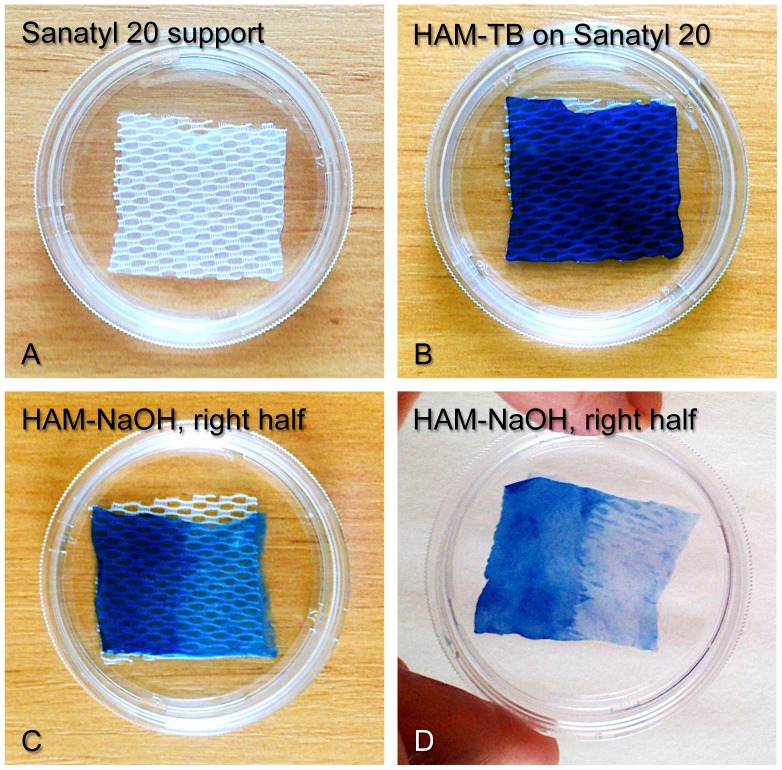
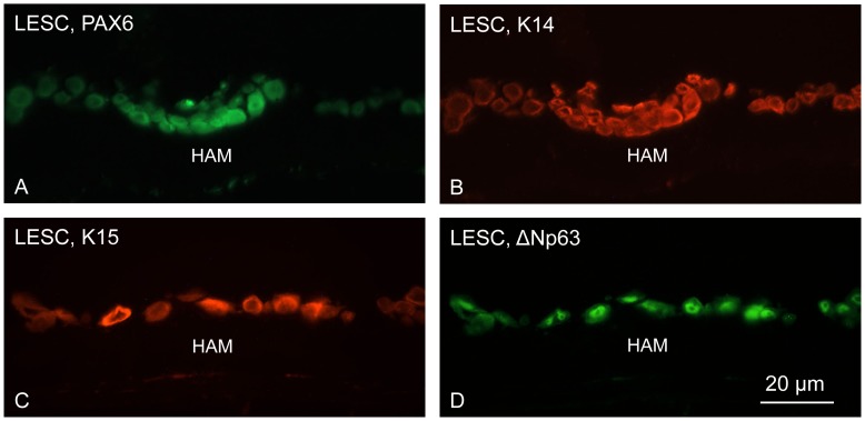
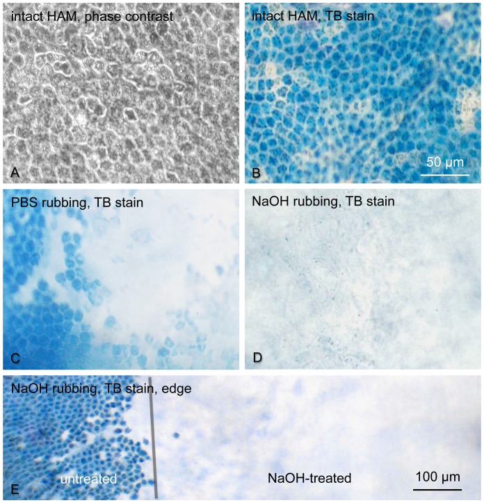
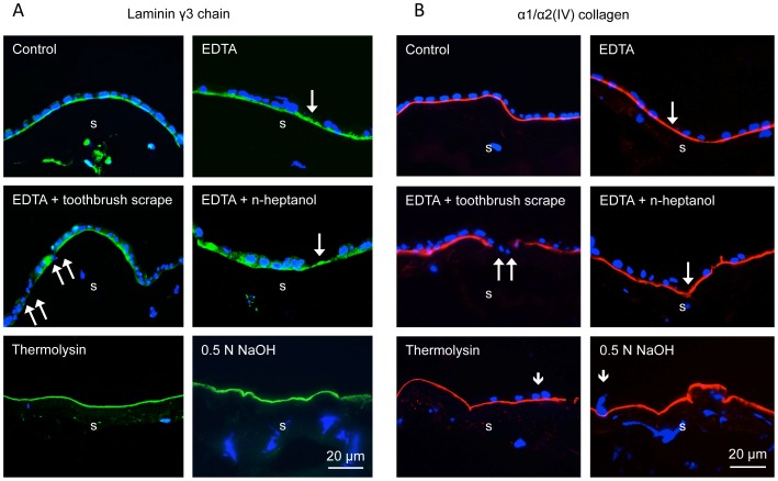
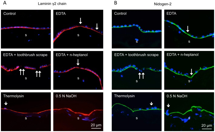
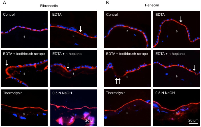
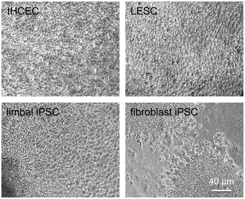
Human amniotic membrane is a standard substratum used to culture limbal epithelial stem cells for transplantation to patients with limbal stem cell deficiency. Various methods were developed to decellularize amniotic membrane, because denuded membrane is poorly immunogenic and better supports repopulation by dissociated limbal epithelial cells. Amniotic membrane denuding usually involves treatment with EDTA and/or proteolytic enzymes; in many cases additional mechanical scraping is required. Although ensuring limbal cell proliferation, these methods are not standardized, require relatively long treatment times and can result in membrane damage. We propose to use 0.5 M NaOH to reliably remove amniotic cells from the membrane. This method was used before to lyse cells for DNA isolation and radioactivity counting. Gently rubbing a cotton swab soaked in NaOH over the epithelial side of amniotic membrane leads to nearly complete and easy removal of adherent cells in less than a minute. The denuded membrane is subsequently washed in a neutral buffer. Cell removal was more thorough and uniform than with EDTA, or EDTA plus mechanical scraping with an electric toothbrush, or n-heptanol plus EDTA treatment. NaOH-denuded amniotic membrane did not show any perforations compared with mechanical or thermolysin denuding, and showed excellent preservation of immunoreactivity for major basement membrane components including laminin α2, γ1-γ3 chains, α1/α2 and α6 type IV collagen chains, fibronectin, nidogen-2, and perlecan. Sodium hydroxide treatment was efficient with fresh or cryopreserved (10% dimethyl sulfoxide or 50% glycerol) amniotic membrane. The latter method is a common way of membrane storage for subsequent grafting in the European Union. NaOH-denuded amniotic membrane supported growth of human limbal epithelial cells, immortalized corneal epithelial cells, and induced pluripotent stem cells. This simple, fast and reliable method can be used to standardize decellularized amniotic membrane preparations for expansion of limbal stem cells in vitro before transplantation to patients.
人羊膜是一种标准的基底用于培养角膜缘上皮干细胞移植到患者的角膜缘干细胞缺乏。各种方法被开发用于脱细胞羊膜,因为裸露的膜是免疫原性差,更好地支持由分离的角膜缘上皮细胞再定植。羊膜脱细胞通常涉及处理 EDTA 和/或蛋白酶;在许多情况下,需要额外的机械刮擦。虽然确保角膜缘细胞增殖,这些方法不标准化,需要相对较长的处理时间,并可能导致膜损伤。我们建议使用 0.5 M NaOH 可靠地从膜中去除羊膜细胞。这种方法以前用于裂解细胞进行 DNA 分离和放射性计数。轻轻地用棉签蘸 NaOH 擦拭羊膜的上皮侧,不到一分钟即可彻底去除几乎所有的黏附细胞。随后用中性缓冲液冲洗裸露的膜。细胞去除比 EDTA 更彻底和均匀,比 EDTA 加电动牙刷机械刮擦或 n-庚醇加 EDTA 处理更彻底和均匀。与机械或糜蛋白酶脱细胞相比,NaOH 脱细胞的羊膜没有任何穿孔,并且显示出对主要基底膜成分的免疫反应性的极好保存,包括层粘连蛋白α2、γ1-γ3 链、α1/α2 和α6 型 IV 胶原链、纤维连接蛋白、巢蛋白-2 和 perlecan。新鲜或冷冻保存(10%二甲基亚砜或 50%甘油)的羊膜对氢氧化钠处理均有效。后一种方法是欧盟中用于随后移植的膜储存的常见方法。NaOH 脱细胞的羊膜支持人角膜缘上皮细胞、永生化角膜上皮细胞和诱导多能干细胞的生长。这种简单、快速和可靠的方法可用于标准化脱细胞羊膜制剂,用于体外扩增角膜缘干细胞,然后移植给患者。