Zendehdel Rezvan, Masoudi-Nejad Ali, Mohammadzadeh Javad, H Shirazi Farshad
Pharmaceutical Research Center, Shahid Beheshti University of Medical Sciences, Tehran, Iran.
Iran J Pharm Res. 2012 Winter;11(1):235-40.
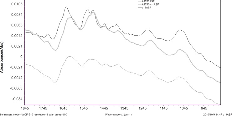
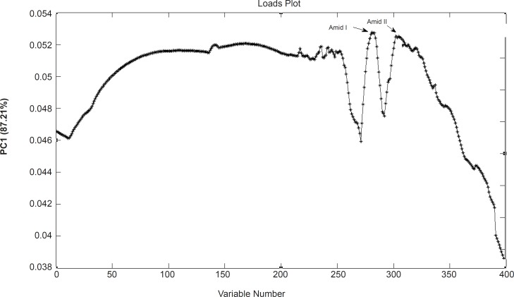
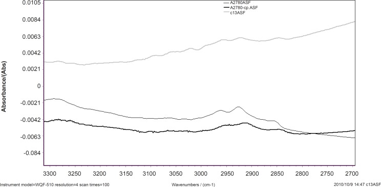
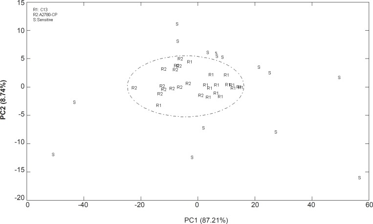
Cisplatin is a common chemotherapeutic agent that used for treatment of many solid cancers. Rapid identification of chemotherapy resistance is very important and may lead to effective treatment plan. Spectroscopy techniques, such as infrared spectroscopy, which are sensitive to biochemical composition of samples, have shown potentials to discriminate tissues. Developing in Fourier transform infrared (FTIR) as a diagnostic tool support conventional technique in investigating cell phenotype. By this goal three different cell lines, two cisplatin resistant OV2008-DDP (C13) and A2780-CP ovarian cell lines and one cisplatin sensitive A2780 cell line were investigated by FTIR spectroscopy. Data were subjected to principle component analysis (PCA) to obtain FTIR pattern for cisplatin resistance. Using FTIR spectroscopy on these cells in the range of 400-4000 cm(-1) was shown dramatic change in cells. Results shows that Cisplatin resistance pattern is characterized in spectrum with the alteration of conformation in secondary structure of proteins and a shift toward the high wave numbers of CH2 stretching vibration. The FTIR data set between 1000 and 3000 cm(-1) could be consumed as biochemical typicality spectra among resistant and sensitive cell lines while correctly classified by PCA model. Our work supports the promise of PCA analysis of FTIR data as a powerful combined approach for the development of automated methods to recognize resistant to cisplatin in experimental cell lines. One of the advantages of this tool is to investigate the resistant percent of cancer cells .Such technique may bring new tool in cancer diagnosis and stage definition in cancerous tissues.
顺铂是一种常用的化疗药物,用于治疗多种实体癌。快速识别化疗耐药性非常重要,可能会带来有效的治疗方案。光谱技术,如对样品生化成分敏感的红外光谱,已显示出区分组织的潜力。开发傅里叶变换红外(FTIR)作为诊断工具,可支持传统技术研究细胞表型。为实现这一目标,通过FTIR光谱研究了三种不同的细胞系,两种顺铂耐药的OV2008-DDP(C13)和A2780-CP卵巢细胞系以及一种顺铂敏感的A2780细胞系。对数据进行主成分分析(PCA)以获得顺铂耐药性的FTIR图谱。在400-4000 cm(-1)范围内对这些细胞使用FTIR光谱显示细胞有显著变化。结果表明,顺铂耐药模式在光谱中表现为蛋白质二级结构构象的改变以及CH2伸缩振动向高波数的偏移。1000至3000 cm(-1)之间的FTIR数据集可作为耐药和敏感细胞系之间的生化典型光谱,同时由PCA模型正确分类。我们的工作支持将FTIR数据的PCA分析作为一种强大的组合方法用于开发自动识别实验细胞系中顺铂耐药性的方法。该工具的优点之一是能够研究癌细胞的耐药百分比。这种技术可能会为癌组织的癌症诊断和分期定义带来新工具。