• 文献检索
  • 文档翻译
  • 深度研究
  • 学术资讯
  • Suppr Zotero 插件Zotero 插件
  • 邀请有礼
  • 套餐&价格
  • 历史记录
应用&插件
Suppr Zotero 插件Zotero 插件浏览器插件Mac 客户端Windows 客户端微信小程序
定价
高级版会员购买积分包购买API积分包
服务
文献检索文档翻译深度研究API 文档MCP 服务
关于我们
关于 Suppr公司介绍联系我们用户协议隐私条款
关注我们

Suppr 超能文献

核心技术专利:CN118964589B侵权必究
粤ICP备2023148730 号-1Suppr @ 2026

文献检索

告别复杂PubMed语法,用中文像聊天一样搜索,搜遍4000万医学文献。AI智能推荐,让科研检索更轻松。

立即免费搜索

文件翻译

保留排版,准确专业,支持PDF/Word/PPT等文件格式,支持 12+语言互译。

免费翻译文档

深度研究

AI帮你快速写综述,25分钟生成高质量综述,智能提取关键信息,辅助科研写作。

立即免费体验

钆塞酸二钠增强肝胆期 MRI 成像在评估局灶性肝脏实性病变中的应用价值。

Added value of Gd-EOB-DTPA-enhanced Hepatobiliary phase MR imaging in evaluation of focal solid hepatic lesions.

机构信息

Department of Radiology, University Medical Center Regensburg, Regensburg 93042, Germany.

出版信息

BMC Med Imaging. 2013 Dec 1;13:41. doi: 10.1186/1471-2342-13-41.

DOI:10.1186/1471-2342-13-41
PMID:24289722
原文链接:https://pmc.ncbi.nlm.nih.gov/articles/PMC3866976/
Abstract

BACKGROUND

Correct characterization of focal solid hepatic lesions has always been a challenge and is of great diagnostic and therapeutic relevance. The purpose of this study was to determine the added value of hepatobiliary phase images in Gd-EOB-DTPA-enhanced magnetic resonance imaging (MRI) for differentiating focal solid hepatic lesions.

METHODS

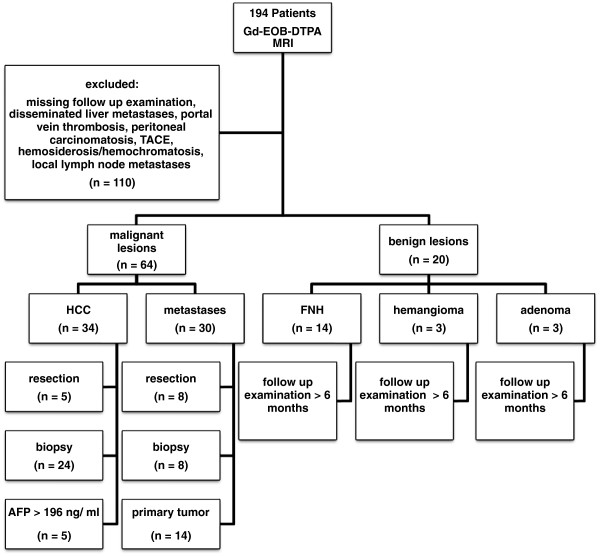
In this retrospective trial 84 consecutive patients underwent Gd-EOB-DTPA-enhanced MR examinations. MRI was conducted for 64 patients with malignant focal hepatic lesions (34 hepatocellular carcinoma (HCC), 30 metastases) and for 20 patients with benign hepatic lesions (14 focal nodular hyperplasia (FNH), 3 adenoma, 3 hemangioma). Five radiologists independently reviewed three sets of MR images by means of a 5-point confidence scale from score 1 (definitely benign) to score 5 (definitely malignant): set 1: unenhanced images; set 2: unenhanced and Gd-EOB-DTPA-enhanced dynamic images; set 3: hepatobiliary phase images in addition to set 2. Accuracy was assessed by the alternative free-response receiver operating characteristic curve (Az) and the index of diagnostic performance was calculated.

RESULTS

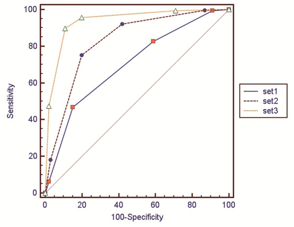
Diagnostic accuracy was significantly improved by the addition of Gd-EOB-DTPA-enhanced dynamic images: Az in set 1 was 0.708 and 0.833 in set 2 (P = 0.0002). The addition of hepatobiliary phase images increased the Az value to 0.941 in set 3 (set 3 vs set 2, P < 0.0001; set 3 vs set 1, P < 0.0001). The index of diagnostic performance was lowest in set 1 (45%), improved in set 2 (71%), and highest in set 3 (94%).

CONCLUSIONS

Hepatobiliary phase images obtained after Gd-EOB-DTPA-enhanced dynamic MRI improve the differentiation of focal solid hepatic lesions.

摘要

背景

准确描述局灶性肝脏实性病变一直是一个挑战,对诊断和治疗具有重要意义。本研究旨在确定钆塞酸二钠增强磁共振成像(MRI)肝胆期成像在鉴别局灶性肝脏实性病变中的附加价值。

方法

本回顾性试验纳入 84 例连续患者行 Gd-EOB-DTPA 增强 MRI 检查。对 64 例恶性局灶性肝脏病变(34 例肝细胞癌(HCC),30 例转移瘤)和 20 例良性肝脏病变(14 例局灶性结节增生(FNH),3 例腺瘤,3 例血管瘤)患者进行 MRI 检查。5 名放射科医生通过 5 分置信度评分(评分 1 表示“肯定良性”,评分 5 表示“肯定恶性”)独立评估三组 MRI 图像:第 1 组:平扫图像;第 2 组:平扫+钆塞酸二钠增强动态图像;第 3 组:第 2 组+肝胆期图像。采用备选自由反应接受者操作特征曲线(Az)评估准确性,并计算诊断性能指标。

结果

加入钆塞酸二钠增强动态图像后,诊断准确性显著提高:第 1 组 Az 为 0.708,第 2 组 Az 为 0.833(P=0.0002)。加入肝胆期图像后,第 3 组 Az 值增加至 0.941(第 3 组与第 2 组相比,P<0.0001;第 3 组与第 1 组相比,P<0.0001)。第 1 组诊断性能指标最低(45%),第 2 组(71%)提高,第 3 组最高(94%)。

结论

钆塞酸二钠增强动态 MRI 后获得的肝胆期图像可改善局灶性肝脏实性病变的鉴别。

https://cdn.ncbi.nlm.nih.gov/pmc/blobs/4df1/3866976/be56108be8e1/1471-2342-13-41-3.jpg
https://cdn.ncbi.nlm.nih.gov/pmc/blobs/4df1/3866976/a6cab22596ae/1471-2342-13-41-1.jpg
https://cdn.ncbi.nlm.nih.gov/pmc/blobs/4df1/3866976/b03ef74405ca/1471-2342-13-41-2.jpg
https://cdn.ncbi.nlm.nih.gov/pmc/blobs/4df1/3866976/be56108be8e1/1471-2342-13-41-3.jpg
https://cdn.ncbi.nlm.nih.gov/pmc/blobs/4df1/3866976/a6cab22596ae/1471-2342-13-41-1.jpg
https://cdn.ncbi.nlm.nih.gov/pmc/blobs/4df1/3866976/b03ef74405ca/1471-2342-13-41-2.jpg
https://cdn.ncbi.nlm.nih.gov/pmc/blobs/4df1/3866976/be56108be8e1/1471-2342-13-41-3.jpg

相似文献

1
Added value of Gd-EOB-DTPA-enhanced Hepatobiliary phase MR imaging in evaluation of focal solid hepatic lesions.钆塞酸二钠增强肝胆期 MRI 成像在评估局灶性肝脏实性病变中的应用价值。
BMC Med Imaging. 2013 Dec 1;13:41. doi: 10.1186/1471-2342-13-41.
2
Value of retrospective fusion of PET and MR images in detection of hepatic metastases: comparison with 18F-FDG PET/CT and Gd-EOB-DTPA-enhanced MRI.回顾性融合 PET 和 MR 图像在肝转移检测中的价值:与 18F-FDG PET/CT 和 Gd-EOB-DTPA 增强 MRI 的比较。
J Nucl Med. 2010 May;51(5):692-9. doi: 10.2967/jnumed.109.068510. Epub 2010 Apr 15.
3
Detection and characterization of focal liver lesions: a Japanese phase III, multicenter comparison between gadoxetic acid disodium-enhanced magnetic resonance imaging and contrast-enhanced computed tomography predominantly in patients with hepatocellular carcinoma and chronic liver disease.检测和描述肝脏局灶性病变:一项日本 III 期、多中心的钆塞酸二钠增强磁共振成像与增强计算机断层扫描对比研究,主要针对肝细胞癌和慢性肝病患者。
Invest Radiol. 2010 Mar;45(3):133-41. doi: 10.1097/RLI.0b013e3181caea5b.
4
Intrapatient Comparison of the Hepatobiliary Phase of Gd-BOPTA and Gd-EOB-DTPA in the Differentiation of Hepatocellular Adenoma From Focal Nodular Hyperplasia.患者内比较 Gd-BOPTA 和 Gd-EOB-DTPA 的肝胆期在肝细胞腺瘤与局灶性结节增生鉴别诊断中的作用。
J Magn Reson Imaging. 2019 Mar;49(3):700-710. doi: 10.1002/jmri.26227. Epub 2018 Sep 25.
5
[Gd-EOB-DTPA-enhanced magnetic resonance imaging: differentiation between focal nodular hyperplasia and hepatocellular adenoma].[钆塞酸二钠增强磁共振成像:局灶性结节性增生与肝细胞腺瘤的鉴别诊断]
Acta Med Port. 2011 Dec;24 Suppl 2:531-8. Epub 2011 Dec 31.
6
Accuracy and confidence of Gd-EOB-DTPA enhanced MRI and diffusion-weighted imaging alone and in combination for the diagnosis of liver metastases.钆塞酸二钠增强 MRI 和弥散加权成像单独及联合应用诊断肝转移瘤的准确性和置信度。
Eur J Radiol. 2013 May;82(5):822-8. doi: 10.1016/j.ejrad.2012.12.005. Epub 2012 Dec 31.
7
Detection and characterisation of focal liver lesions in colorectal carcinoma patients: comparison of diffusion-weighted and Gd-EOB-DTPA enhanced MR imaging.检测和特征分析结直肠癌患者肝内局灶性病变:弥散加权成像与钆塞酸二钠增强磁共振成像的对比研究。
Eur Radiol. 2011 Apr;21(4):832-40. doi: 10.1007/s00330-010-1977-2. Epub 2010 Oct 2.
8
Washout appearance in Gd-EOB-DTPA-enhanced MR imaging: A differentiating feature between hepatocellular carcinoma with paradoxical uptake on the hepatobiliary phase and focal nodular hyperplasia-like nodules.钆塞酸二钠增强磁共振成像中的廓清表现:肝胆期呈反向摄取的肝细胞癌与局灶性结节样增生样结节之间的鉴别特征
J Magn Reson Imaging. 2017 Jun;45(6):1599-1608. doi: 10.1002/jmri.25493. Epub 2016 Oct 11.
9
Imaging features of intrahepatic cholangiocarcinoma in Gd-EOB-DTPA-enhanced MRI.钆塞酸二钠增强 MRI 成像对肝内胆管细胞癌的影像学特征。
Eur J Radiol. 2013 Mar;82(3):e101-6. doi: 10.1016/j.ejrad.2012.10.010. Epub 2012 Nov 16.
10
Gd-EOB-DTPA-enhanced magnetic resonance imaging features of hepatic hemangioma compared with enhanced computed tomography.钆塞酸二钠增强磁共振成像与增强 CT 对肝血管瘤的影像学特征比较。
World J Gastroenterol. 2012 Nov 21;18(43):6269-76. doi: 10.3748/wjg.v18.i43.6269.

引用本文的文献

1
High-precision MRI of liver and hepatic lesions on gadoxetic acid-enhanced hepatobiliary phase using a deep learning technique.使用深度学习技术对钆塞酸增强肝胆期肝脏及肝脏病变进行高精度磁共振成像。
Jpn J Radiol. 2025 Apr;43(4):649-655. doi: 10.1007/s11604-024-01693-2. Epub 2024 Nov 11.
2
[Application of Diffusion-Weighted Imaging and Hepatobiliary-Specific Contrast Agent Gd-EOB-DTPA in the Diagnosis and Differential Diagnosis of Focal Liver Lesions].扩散加权成像及肝胆特异性对比剂钆塞酸二钠在肝脏局灶性病变诊断及鉴别诊断中的应用
Sichuan Da Xue Xue Bao Yi Xue Ban. 2022 Sep;53(5):737-743. doi: 10.12182/20220960205.
3

本文引用的文献

1
Hepatocellular carcinoma: imaging patterns on gadoxetic acid-enhanced MR Images and their value as an imaging biomarker.肝细胞癌:钆塞酸增强磁共振成像的影像学表现及其作为影像学生物标志物的价值。
Radiology. 2013 Jun;267(3):776-86. doi: 10.1148/radiol.13120775. Epub 2013 Feb 11.
2
Diagnostic efficacy of gadoxetic acid-enhanced MRI for the detection and characterisation of liver metastases: comparison with multidetector-row CT.钆塞酸增强 MRI 检测和特征分析肝脏转移瘤的诊断效能:与多排 CT 的对比研究。
Br J Radiol. 2012 May;85(1013):539-47. doi: 10.1259/bjr/25139667.
3
Hepatocellular adenoma and focal nodular hyperplasia: value of gadoxetic acid-enhanced MR imaging in differential diagnosis.
Quantitative analysis of liver function: 3D variable-flip-angle versus Look-Locker T1 relaxometry in hepatocyte-specific contrast-enhanced liver MRI.
肝功能定量分析:肝细胞特异性对比增强肝脏磁共振成像中3D可变翻转角与Look-Locker T1弛豫测量法的比较
Quant Imaging Med Surg. 2022 Apr;12(4):2509-2522. doi: 10.21037/qims-21-597.
4
MRI characterization of focal liver lesions in non-cirrhotic patients: assessment of added value of gadoxetic acid-enhanced hepatobiliary phase imaging.非肝硬化患者肝脏局灶性病变的MRI特征:钆塞酸增强肝胆期成像附加值的评估
Insights Imaging. 2020 Sep 22;11(1):101. doi: 10.1186/s13244-020-00894-3.
5
Hepatic tumors: pitfall in diagnostic imaging.肝脏肿瘤:诊断影像学中的陷阱。
Acta Biomed. 2020 Jul 13;91(8-S):9-17. doi: 10.23750/abm.v91i8-S.9969.
6
A CT-based radiomics nomogram for differentiation of focal nodular hyperplasia from hepatocellular carcinoma in the non-cirrhotic liver.基于 CT 的放射组学列线图用于区分非肝硬化肝脏中的局灶性结节性增生与肝细胞癌。
Cancer Imaging. 2020 Feb 24;20(1):20. doi: 10.1186/s40644-020-00297-z.
7
Improved visualisation of hepatic metastases in gadoxetate disodium-enhanced MRI: Potential of contrast-optimised (phase-sensitive) inversion recovery imaging.钆塞酸二钠增强 MRI 中肝转移瘤的可视化改善:对比优化(相位敏感)反转恢复成像的潜力。
PLoS One. 2019 Mar 6;14(3):e0213408. doi: 10.1371/journal.pone.0213408. eCollection 2019.
8
Comparison of values of CT and MRI imaging in the diagnosis of hepatocellular carcinoma and analysis of prognostic factors.CT与MRI成像在肝细胞癌诊断中的价值比较及预后因素分析
Oncol Lett. 2019 Jan;17(1):1184-1188. doi: 10.3892/ol.2018.9690. Epub 2018 Nov 12.
9
Functional imaging of hepatocellular carcinoma.肝细胞癌的功能成像
Hepat Oncol. 2016 Apr;3(2):137-153. doi: 10.2217/hep-2015-0005. Epub 2016 Mar 29.
10
Extracellular contrast agent-enhanced MRI: 15-min delayed phase may improve the diagnostic performance for hepatocellular carcinoma in patients with chronic liver disease.细胞外对比剂增强 MRI:15 分钟延迟期可提高慢性肝病患者肝细胞癌的诊断性能。
Eur Radiol. 2018 Apr;28(4):1551-1559. doi: 10.1007/s00330-017-5119-y. Epub 2017 Nov 13.
肝细胞腺瘤和局灶性结节性增生:钆塞酸增强磁共振成像在鉴别诊断中的价值。
Radiology. 2012 Feb;262(2):520-9. doi: 10.1148/radiol.11101742.
4
Fate of hypointense lesions on Gd-EOB-DTPA-enhanced magnetic resonance imaging.钆塞酸二钠增强磁共振成像上低信号病灶的转归。
Eur J Radiol. 2012 Nov;81(11):2973-7. doi: 10.1016/j.ejrad.2012.01.007. Epub 2012 Jan 26.
5
Small (≤ 2 cm) hepatocellular carcinoma in patients with chronic liver disease: comparison of gadoxetic acid-enhanced 3.0 T MRI and multiphasic 64-multirow detector CT.慢性肝病患者小肝癌(≤ 2cm):钆塞酸增强 3.0T MRI 与多期 64 排多层螺旋 CT 的对比研究。
Br J Radiol. 2012 Jul;85(1015):e314-22. doi: 10.1259/bjr/27727228. Epub 2011 Dec 13.
6
Imaging study of early hepatocellular carcinoma: usefulness of gadoxetic acid-enhanced MR imaging.早期肝细胞癌的影像学研究:钆塞酸增强磁共振成像的作用。
Radiology. 2011 Dec;261(3):834-44. doi: 10.1148/radiol.11101840. Epub 2011 Oct 13.
7
Added value of gadoxetic acid-enhanced hepatobiliary phase MR imaging in the diagnosis of hepatocellular carcinoma.钆塞酸增强肝胆期磁共振成像在肝细胞癌诊断中的应用价值。
Radiology. 2010 May;255(2):459-66. doi: 10.1148/radiol.10091388.
8
Detection and characterization of focal liver lesions: a Japanese phase III, multicenter comparison between gadoxetic acid disodium-enhanced magnetic resonance imaging and contrast-enhanced computed tomography predominantly in patients with hepatocellular carcinoma and chronic liver disease.检测和描述肝脏局灶性病变:一项日本 III 期、多中心的钆塞酸二钠增强磁共振成像与增强计算机断层扫描对比研究,主要针对肝细胞癌和慢性肝病患者。
Invest Radiol. 2010 Mar;45(3):133-41. doi: 10.1097/RLI.0b013e3181caea5b.
9
Gadoxetic acid-enhanced magnetic resonance imaging for differentiating small hepatocellular carcinomas (< or =2 cm in diameter) from arterial enhancing pseudolesions: special emphasis on hepatobiliary phase imaging.钆塞酸增强磁共振成像在鉴别 < 或 = 2cm 直径的小肝癌和动脉期增强假性病灶中的应用:特别强调肝胆期成像。
Invest Radiol. 2010 Feb;45(2):96-103. doi: 10.1097/RLI.0b013e3181c5faf7.
10
Consensus report of the 2nd International Forum for Liver MRI.第二届国际肝脏磁共振成像论坛共识报告
Eur Radiol. 2009 Oct;19 Suppl 5:S975-89. doi: 10.1007/s00330-009-1624-y.