• 文献检索
  • 文档翻译
  • 深度研究
  • 学术资讯
  • Suppr Zotero 插件Zotero 插件
  • 邀请有礼
  • 套餐&价格
  • 历史记录
应用&插件
Suppr Zotero 插件Zotero 插件浏览器插件Mac 客户端Windows 客户端微信小程序
定价
高级版会员购买积分包购买API积分包
服务
文献检索文档翻译深度研究API 文档MCP 服务
关于我们
关于 Suppr公司介绍联系我们用户协议隐私条款
关注我们

Suppr 超能文献

核心技术专利:CN118964589B侵权必究
粤ICP备2023148730 号-1Suppr @ 2026

文献检索

告别复杂PubMed语法,用中文像聊天一样搜索,搜遍4000万医学文献。AI智能推荐,让科研检索更轻松。

立即免费搜索

文件翻译

保留排版,准确专业,支持PDF/Word/PPT等文件格式,支持 12+语言互译。

免费翻译文档

深度研究

AI帮你快速写综述,25分钟生成高质量综述,智能提取关键信息,辅助科研写作。

立即免费体验

基于 CT 的放射组学列线图用于区分非肝硬化肝脏中的局灶性结节性增生与肝细胞癌。

A CT-based radiomics nomogram for differentiation of focal nodular hyperplasia from hepatocellular carcinoma in the non-cirrhotic liver.

机构信息

Department of Radiology, the Affiliated Hospital of Qingdao University, No.16, Jiangsu Road, Qingdao, 266000, Shandong, China.

Department of Nuclear Medicine, the Affiliated Hospital of Qingdao University, Qingdao, Shandong, China.

出版信息

Cancer Imaging. 2020 Feb 24;20(1):20. doi: 10.1186/s40644-020-00297-z.

DOI:10.1186/s40644-020-00297-z
PMID:32093786
原文链接:https://pmc.ncbi.nlm.nih.gov/articles/PMC7041197/
Abstract

BACKGROUND

The purpose of this study was to develop and validate a radiomics nomogram for preoperative differentiating focal nodular hyperplasia (FNH) from hepatocellular carcinoma (HCC) in the non-cirrhotic liver.

METHODS

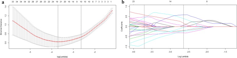
A total of 156 patients with FNH (n = 55) and HCC (n = 101) were divided into a training set (n = 119) and a validation set (n = 37). Radiomics features were extracted from triphasic contrast CT images. A radiomics signature was constructed with the least absolute shrinkage and selection operator algorithm, and a radiomics score (Rad-score) was calculated. Clinical data and CT findings were assessed to build a clinical factors model. Combined with the Rad-score and independent clinical factors, a radiomics nomogram was constructed by multivariate logistic regression analysis. Nomogram performance was assessed with respect to discrimination and clinical usefulness.

RESULTS

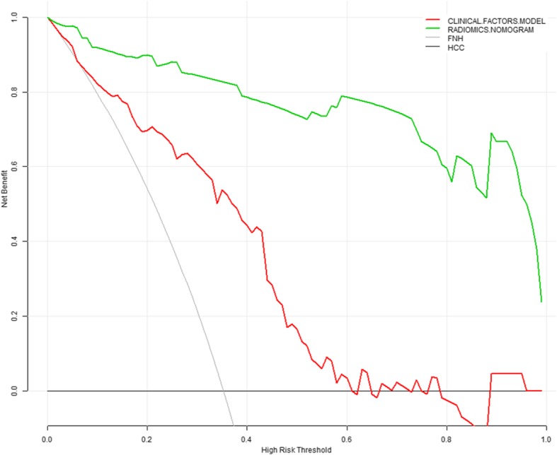
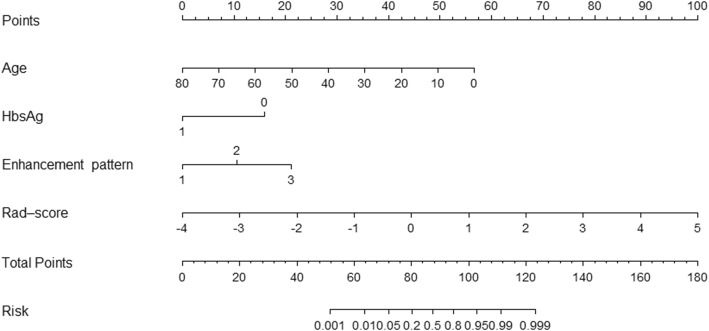
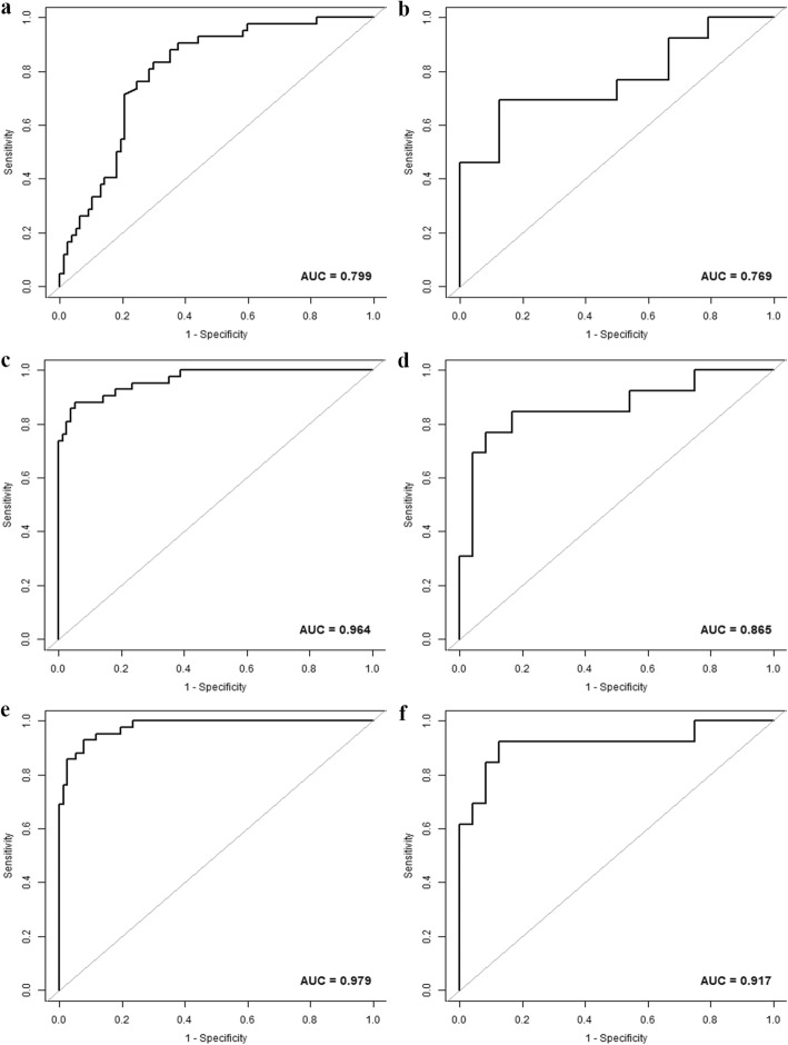
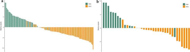
Four thousand two hundred twenty-seven features were extracted and reduced to 10 features as the most important discriminators to build the radiomics signature. The radiomics signature showed good discrimination in the training set (AUC [area under the curve], 0.964; 95% confidence interval [CI], 0.934-0.995) and the validation set (AUC, 0.865; 95% CI, 0.725-1.000). Age, Hepatitis B virus infection, and enhancement pattern were the independent clinical factors. The radiomics nomogram, which incorporated the Rad-score and clinical factors, showed good discrimination in the training set (AUC, 0.979; 95% CI, 0.959-0.998) and the validation set (AUC, 0.917; 95% CI, 0.800-1.000), and showed better discrimination capability (P < 0.001) compared with the clinical factors model (AUC, 0.799; 95% CI, 0.719-0.879) in the training set. Decision curve analysis showed the nomogram outperformed the clinical factors model in terms of clinical usefulness.

CONCLUSIONS

The CT-based radiomics nomogram, a noninvasive preoperative prediction tool that incorporates the Rad-score and clinical factors, shows favorable predictive efficacy for differentiating FNH from HCC in the non-cirrhotic liver, which might facilitate clinical decision-making process.

摘要

背景

本研究旨在建立并验证一个针对非肝硬化肝脏局灶性结节性增生(FNH)与肝细胞癌(HCC)的术前鉴别诊断的影像组学列线图。

方法

共纳入 156 例 FNH(n=55)和 HCC(n=101)患者,分为训练集(n=119)和验证集(n=37)。从三期增强 CT 图像中提取影像组学特征。采用最小绝对值收缩和选择算子算法构建影像组学特征签名,并计算影像组学评分(Rad-score)。评估临床数据和 CT 表现以构建临床因素模型。通过多变量逻辑回归分析,结合 Rad-score 和独立临床因素,构建影像组学列线图。采用判别效能和临床实用性评估列线图的性能。

结果

提取 4227 个特征,降维至 10 个最重要的鉴别特征,构建影像组学特征签名。该影像组学特征在训练集(AUC [曲线下面积],0.964;95%置信区间 [CI],0.934-0.995)和验证集(AUC,0.865;95%CI,0.725-1.000)中均具有良好的判别能力。年龄、乙型肝炎病毒感染和增强模式是独立的临床因素。纳入 Rad-score 和临床因素的影像组学列线图在训练集(AUC,0.979;95%CI,0.959-0.998)和验证集(AUC,0.917;95%CI,0.800-1.000)中具有良好的判别能力,与训练集的临床因素模型(AUC,0.799;95%CI,0.719-0.879)相比,具有更好的判别能力(P<0.001)。决策曲线分析显示,该列线图在临床实用性方面优于临床因素模型。

结论

该基于 CT 的影像组学列线图是一种非侵入性的术前预测工具,纳入了 Rad-score 和临床因素,对于鉴别非肝硬化肝脏中的 FNH 和 HCC 具有良好的预测效能,有助于临床决策过程。

https://cdn.ncbi.nlm.nih.gov/pmc/blobs/2972/7041197/6b54036eeafd/40644_2020_297_Fig5_HTML.jpg
https://cdn.ncbi.nlm.nih.gov/pmc/blobs/2972/7041197/53ba5251045a/40644_2020_297_Fig1_HTML.jpg
https://cdn.ncbi.nlm.nih.gov/pmc/blobs/2972/7041197/24022df8138f/40644_2020_297_Fig2_HTML.jpg
https://cdn.ncbi.nlm.nih.gov/pmc/blobs/2972/7041197/681b31413175/40644_2020_297_Fig3_HTML.jpg
https://cdn.ncbi.nlm.nih.gov/pmc/blobs/2972/7041197/e17d4ceea7f0/40644_2020_297_Fig4_HTML.jpg
https://cdn.ncbi.nlm.nih.gov/pmc/blobs/2972/7041197/6b54036eeafd/40644_2020_297_Fig5_HTML.jpg

相似文献

1
A CT-based radiomics nomogram for differentiation of focal nodular hyperplasia from hepatocellular carcinoma in the non-cirrhotic liver.基于 CT 的放射组学列线图用于区分非肝硬化肝脏中的局灶性结节性增生与肝细胞癌。
Cancer Imaging. 2020 Feb 24;20(1):20. doi: 10.1186/s40644-020-00297-z.
2
CT-Based Radiomics Nomogram: A Potential Tool for Differentiating Hepatocellular Adenoma From Hepatocellular Carcinoma in the Noncirrhotic Liver.基于 CT 的放射组学列线图:一种在非肝硬化肝脏中鉴别肝细胞腺瘤与肝细胞癌的潜在工具。
Acad Radiol. 2021 Jun;28(6):799-807. doi: 10.1016/j.acra.2020.04.027. Epub 2020 May 5.
3
A CT-based radiomics nomogram for differentiation of renal angiomyolipoma without visible fat from homogeneous clear cell renal cell carcinoma.基于 CT 的影像组学列线图,用于区分无可见脂肪的肾血管平滑肌脂肪瘤与均质透明细胞肾细胞癌。
Eur Radiol. 2020 Feb;30(2):1274-1284. doi: 10.1007/s00330-019-06427-x. Epub 2019 Sep 10.
4
A radiomics nomogram for preoperative prediction of microvascular invasion risk in hepatitis B virus-related hepatocellular carcinoma.一种用于术前预测乙型肝炎病毒相关肝细胞癌微血管侵犯风险的影像组学列线图。
Diagn Interv Radiol. 2018 May-Jun;24(3):121-127. doi: 10.5152/dir.2018.17467.
5
A radiomics-based nomogram for the preoperative prediction of posthepatectomy liver failure in patients with hepatocellular carcinoma.一种基于影像组学的列线图,用于术前预测肝细胞癌患者肝切除术后肝衰竭。
Surg Oncol. 2019 Mar;28:78-85. doi: 10.1016/j.suronc.2018.11.013. Epub 2018 Nov 14.
6
F-FDG PET/CT-based radiomics nomogram for preoperative prediction of macrotrabecular-massive hepatocellular carcinoma: a two-center study.基于F-FDG PET/CT的影像组学列线图用于术前预测大结节型-巨块型肝细胞癌:一项双中心研究
Abdom Radiol (NY). 2023 Feb;48(2):532-542. doi: 10.1007/s00261-022-03722-y. Epub 2022 Nov 12.
7
An MR-based radiomics model for differentiation between hepatocellular carcinoma and focal nodular hyperplasia in non-cirrhotic liver.基于磁共振的放射组学模型在非肝硬化肝脏中鉴别肝细胞癌和局灶性结节增生
World J Surg Oncol. 2021 Jun 21;19(1):181. doi: 10.1186/s12957-021-02266-7.
8
Development of a computed tomography-based radiomics nomogram for prediction of transarterial chemoembolization refractoriness in hepatocellular carcinoma.基于计算机断层扫描的影像组学列线图用于预测肝细胞癌经动脉化疗栓塞难治性的研究进展
World J Gastroenterol. 2021 Jan 14;27(2):189-207. doi: 10.3748/wjg.v27.i2.189.
9
A Radiomics Nomogram for the Preoperative Prediction of Lymph Node Metastasis in Bladder Cancer.膀胱癌淋巴结转移术前预测的放射组学列线图
Clin Cancer Res. 2017 Nov 15;23(22):6904-6911. doi: 10.1158/1078-0432.CCR-17-1510. Epub 2017 Sep 5.
10
A radiomics nomogram for the prediction of overall survival in patients with hepatocellular carcinoma after hepatectomy.基于影像组学的Nomogram 模型预测肝癌患者肝切除术后的总生存情况
Cancer Imaging. 2020 Nov 16;20(1):82. doi: 10.1186/s40644-020-00360-9.

引用本文的文献

1
A T2 weighted imaging-based radiomics nomogram for the classification of hepatic blood-rich lesions: hepatocellular carcinoma and benign liver lesions.基于T2加权成像的放射组学列线图用于肝血供丰富病变的分类:肝细胞癌和良性肝病变
Discov Oncol. 2025 Jul 1;16(1):1223. doi: 10.1007/s12672-025-02868-7.
2
Role of AI in empowering and redefining the oncology care landscape: perspective from a developing nation.人工智能在赋能和重新定义肿瘤护理格局中的作用:来自一个发展中国家的视角。
Front Digit Health. 2025 Mar 4;7:1550407. doi: 10.3389/fdgth.2025.1550407. eCollection 2025.
3
Development and validation of an ultrasound-based interpretable machine learning model for the classification of ≤3 cm hepatocellular carcinoma: a multicentre retrospective diagnostic study.

本文引用的文献

1
Hepatocellular Carcinoma: Texture Analysis of Preoperative Computed Tomography Images Can Provide Markers of Tumor Grade and Disease-Free Survival.肝细胞癌:术前 CT 图像纹理分析可为肿瘤分级和无病生存提供标志物。
Korean J Radiol. 2019 Apr;20(4):569-579. doi: 10.3348/kjr.2018.0501.
2
Radiomic analysis of contrast-enhanced CT predicts microvascular invasion and outcome in hepatocellular carcinoma.基于增强 CT 的影像组学分析预测肝细胞癌的微血管侵犯及预后
J Hepatol. 2019 Jun;70(6):1133-1144. doi: 10.1016/j.jhep.2019.02.023. Epub 2019 Mar 13.
3
Radiomics-based classification of hepatocellular carcinoma and hepatic haemangioma on precontrast magnetic resonance images.
基于超声的≤3cm肝细胞癌分类可解释机器学习模型的开发与验证:一项多中心回顾性诊断研究
EClinicalMedicine. 2025 Feb 13;81:103098. doi: 10.1016/j.eclinm.2025.103098. eCollection 2025 Mar.
4
The role of artificial intelligence and image processing in the diagnosis, treatment, and prognosis of liver cancer: a narrative-review.人工智能与图像处理在肝癌诊断、治疗及预后中的作用:一项叙述性综述。
Prz Gastroenterol. 2024;19(3):221-230. doi: 10.5114/pg.2024.143147. Epub 2024 Sep 18.
5
Systematic review and meta-analysis on the classification metrics of machine learning algorithm based radiomics in hepatocellular carcinoma diagnosis.基于机器学习算法的放射组学在肝细胞癌诊断中分类指标的系统评价与荟萃分析
Heliyon. 2024 Aug 14;10(16):e36313. doi: 10.1016/j.heliyon.2024.e36313. eCollection 2024 Aug 30.
6
Prediction models for differentiating benign from malignant liver lesions based on multiparametric dual-energy non-contrast CT.基于多参数双能量非增强CT鉴别肝脏良恶性病变的预测模型
Eur Radiol. 2025 Mar;35(3):1361-1377. doi: 10.1007/s00330-024-11024-8. Epub 2024 Aug 26.
7
Gd-EOB-DTPA enhanced MRI nomogram model to differentiate hepatocellular carcinoma and focal nodular hyperplasia both showing iso- or hyperintensity in the hepatobiliary phase.钆塞酸二钠增强 MRI 列线图模型鉴别肝胆期呈等或高信号的肝细胞肝癌和局灶性结节增生。
BMC Med Imaging. 2024 Aug 12;24(1):211. doi: 10.1186/s12880-024-01382-6.
8
Radiomics-Based Classification of Tumor and Healthy Liver on Computed Tomography Images.基于放射组学的计算机断层扫描图像上肿瘤与健康肝脏的分类
Cancers (Basel). 2024 Mar 14;16(6):1158. doi: 10.3390/cancers16061158.
9
Radiomics nomogram for preoperative differentiation of early-stage serous borderline ovarian tumors and serous malignant ovarian tumors.用于早期浆液性交界性卵巢肿瘤和浆液性恶性卵巢肿瘤术前鉴别的影像组学列线图。
Front Oncol. 2024 Jan 15;13:1269589. doi: 10.3389/fonc.2023.1269589. eCollection 2023.
10
Case Report: The value of contrast-enhanced ultrasound and contrast-enhanced computed tomography in the diagnosis of hepatic angiosarcoma.病例报告:超声造影和增强计算机断层扫描在肝血管肉瘤诊断中的价值。
Front Oncol. 2023 Dec 1;13:1283544. doi: 10.3389/fonc.2023.1283544. eCollection 2023.
基于影像组学的肝细胞癌和肝血管瘤在对比前磁共振图像上的分类
BMC Med Imaging. 2019 Mar 11;19(1):23. doi: 10.1186/s12880-019-0321-9.
4
A radiomics-based nomogram for the preoperative prediction of posthepatectomy liver failure in patients with hepatocellular carcinoma.一种基于影像组学的列线图,用于术前预测肝细胞癌患者肝切除术后肝衰竭。
Surg Oncol. 2019 Mar;28:78-85. doi: 10.1016/j.suronc.2018.11.013. Epub 2018 Nov 14.
5
CT-based peritumoral radiomics signatures to predict early recurrence in hepatocellular carcinoma after curative tumor resection or ablation.基于 CT 的肿瘤周围放射组学特征预测肝癌根治性切除或消融术后早期复发。
Cancer Imaging. 2019 Feb 27;19(1):11. doi: 10.1186/s40644-019-0197-5.
6
Radiomics on Gadoxetic Acid-Enhanced Magnetic Resonance Imaging for Prediction of Postoperative Early and Late Recurrence of Single Hepatocellular Carcinoma.钆塞酸二钠增强磁共振成像的影像组学用于预测单发性肝细胞癌术后早期和晚期复发
Clin Cancer Res. 2019 Jul 1;25(13):3847-3855. doi: 10.1158/1078-0432.CCR-18-2861. Epub 2019 Feb 26.
7
Preoperative radiomics nomogram for microvascular invasion prediction in hepatocellular carcinoma using contrast-enhanced CT.基于增强 CT 的术前影像组学Nomogram 模型预测肝细胞癌微血管侵犯
Eur Radiol. 2019 Jul;29(7):3595-3605. doi: 10.1007/s00330-018-5985-y. Epub 2019 Feb 15.
8
MRI texture analysis for differentiation of malignant and benign hepatocellular tumors in the non-cirrhotic liver.非肝硬化肝脏中恶性与良性肝细胞肿瘤鉴别的MRI纹理分析
Heliyon. 2018 Nov 30;4(11):e00987. doi: 10.1016/j.heliyon.2018.e00987. eCollection 2018 Nov.
9
Volumetric quantitative histogram analysis using diffusion-weighted magnetic resonance imaging to differentiate HCC from other primary liver cancers.利用扩散加权磁共振成像进行容积定量直方图分析以鉴别 HCC 与其他原发性肝癌。
Abdom Radiol (NY). 2019 Mar;44(3):912-922. doi: 10.1007/s00261-019-01906-7.
10
Radiogenomics and Radiomics in Liver Cancers.肝癌中的放射基因组学和放射组学
Diagnostics (Basel). 2018 Dec 27;9(1):4. doi: 10.3390/diagnostics9010004.