Arthritis Res Ther. 2013 Oct 9;15(5):R150. doi: 10.1186/ar4333.
Our recent study indicated that subchondral bone pathogenesis in osteoarthritis (OA) is associated with osteocyte morphology and phenotypic abnormalities. However, the mechanism underlying this abnormality needs to be identified. In this study we investigated the effect of extracellular matrix (ECM) produced from normal and OA bone on osteocytic cells function.
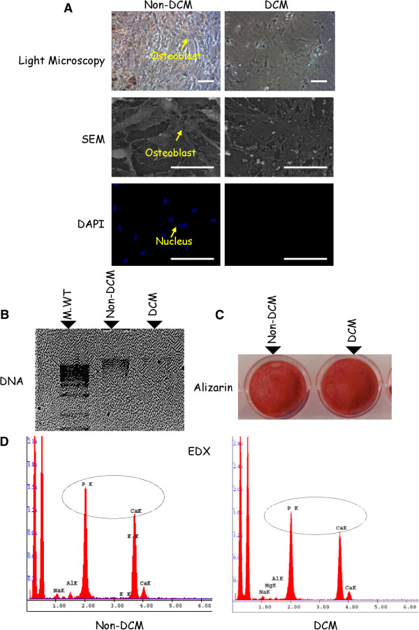
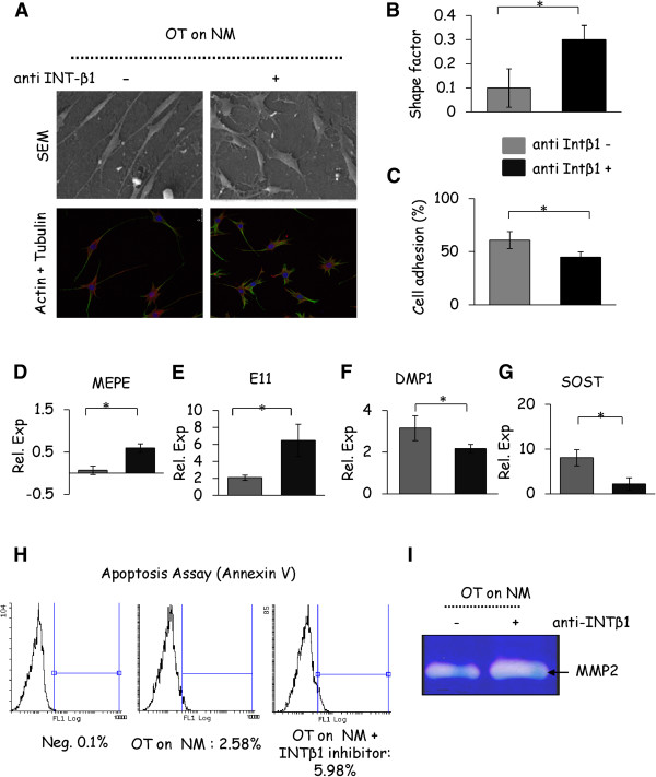
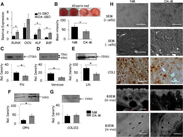
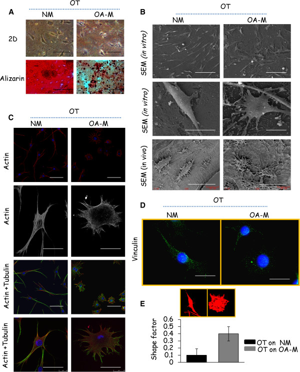
De-cellularized matrices, resembling the bone provisional ECM secreted from primary human subchondral bone osteoblasts (SBOs) of normal and OA patients were used as a model to study the effect on osteocytic cells. Osteocytic cells (MLOY4 osteocyte cell line) cultured on normal and OA derived ECMs were analyzed by confocal microscopy, scanning electron microscopy (SEM), cell attachment assays, zymography, apoptosis assays, qRT-PCR and western blotting. The role of integrinβ1 and focal adhesion kinase (FAK) signaling pathways during these interactions were monitored using appropriate blocking antibodies.
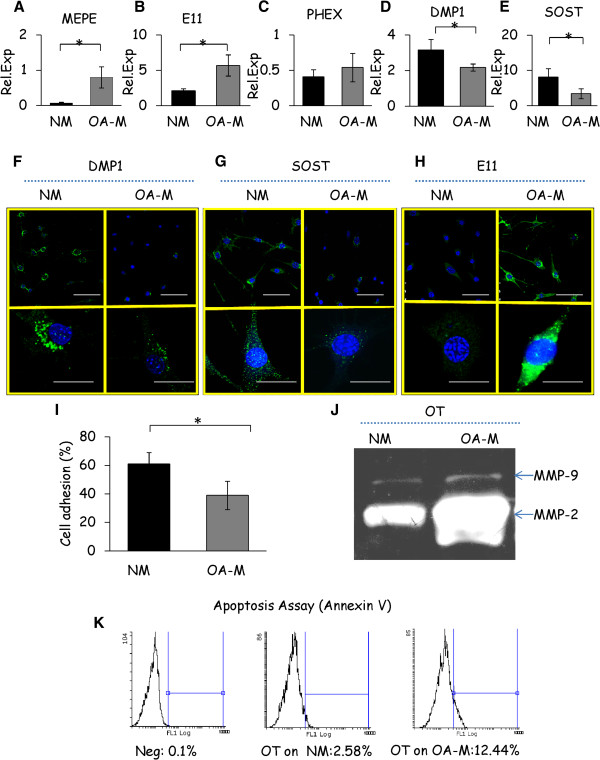
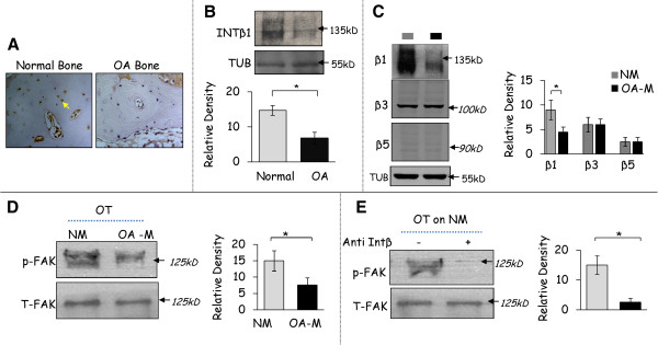
The ECM produced by OA SBOs contained less mineral content, showed altered organization of matrix proteins and matrix structure compared with the matrices produced by normal SBOs. Culture of osteocytic cells on these defective OA ECM resulted in a decrease of integrinβ1 expression and the de-activation of FAK cell signaling pathway, which subsequently affected the initial osteocytic cell's attachment and functions including morphological abnormalities of cytoskeletal structures, focal adhesions, increased apoptosis, altered osteocyte specific gene expression and increased Matrix metalloproteinases (MMP-2) and -9 expression.
This study provides new insights in understanding how altered OA bone matrix can lead to the abnormal osteocyte phenotypic changes, which is typical in OA pathogenesis.
我们最近的研究表明,骨关节炎(OA)中的软骨下骨发病机制与骨细胞形态和表型异常有关。然而,这种异常的机制仍需要确定。在这项研究中,我们研究了来自正常和 OA 骨的细胞外基质(ECM)对骨细胞功能的影响。
去细胞基质类似于正常和 OA 患者的原发性人软骨下骨成骨细胞(SBO)分泌的骨临时 ECM,可作为研究骨细胞的模型。在正常和 OA 衍生的 ECM 上培养的骨细胞(MLOY4 成骨细胞系)通过共聚焦显微镜、扫描电子显微镜(SEM)、细胞附着测定、明胶酶谱分析、凋亡测定、qRT-PCR 和 Western blot 进行分析。使用适当的阻断抗体监测整合素β1 和粘着斑激酶(FAK)信号通路在这些相互作用中的作用。
与正常 SBO 产生的基质相比,OA SBO 产生的 ECM 中矿物质含量较少,基质蛋白和基质结构的组织发生改变。在这些有缺陷的 OA ECM 上培养骨细胞会导致整合素β1表达减少和 FAK 细胞信号通路失活,从而影响初始骨细胞的附着和功能,包括细胞骨架结构、粘着斑的形态异常、凋亡增加、骨细胞特异性基因表达改变以及基质金属蛋白酶(MMP-2)和 -9 表达增加。
这项研究为理解 OA 骨基质如何导致异常的骨细胞表型变化提供了新的见解,这是 OA 发病机制中的典型特征。