Department of Biochemistry and Biophysics, Perelman School of Medicine University of Pennsylvania, Philadelphia, Pennsylvania, United States of America.
PLoS One. 2013 Nov 26;8(11):e77758. doi: 10.1371/journal.pone.0077758. eCollection 2013.
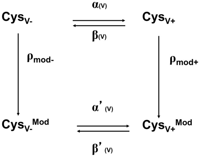
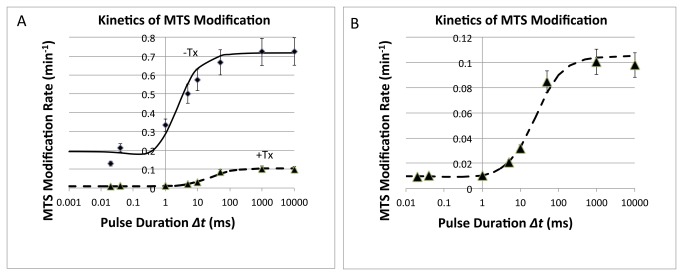
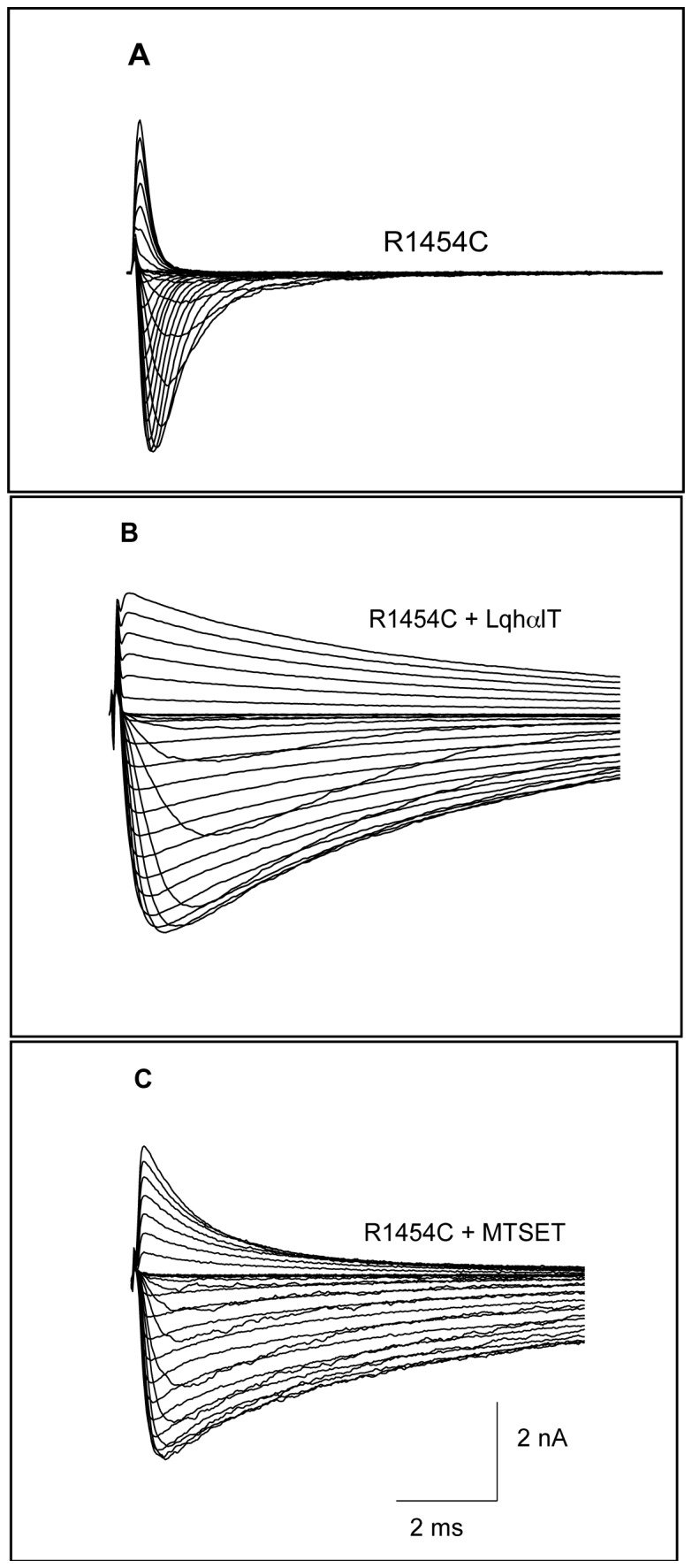
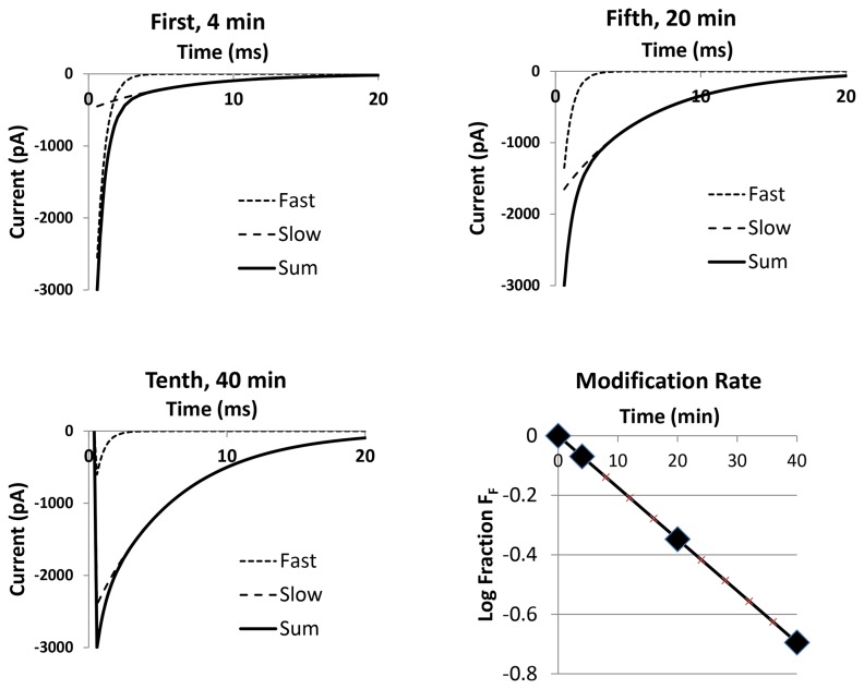
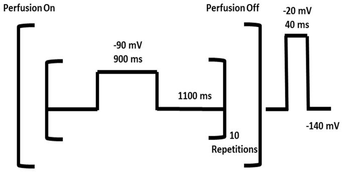
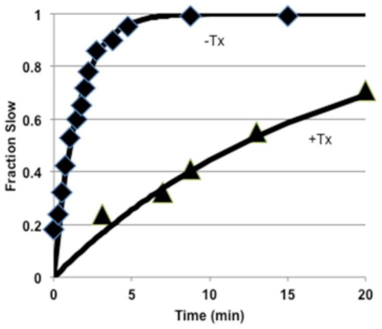
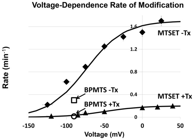
The position of the voltage-sensing transmembrane segment, S4, in voltage-gated ion channels as a function of voltage remains incompletely elucidated. Site-3 toxins bind primarily to the extracellular loops connecting transmembrane helical segments S1-S2 and S3-S4 in Domain 4 (D4) and S5-S6 in Domain 1 (D1) and slow fast-inactivation of voltage-gated sodium channels. As S4 of the human skeletal muscle voltage-gated sodium channel, hNav1.4, moves in response to depolarization from the resting to the inactivated state, two D4S4 reporters (R2C and R3C, Arg1451Cys and Arg1454Cys, respectively) move from internal to external positions as deduced by reactivity to internally or externally applied sulfhydryl group reagents, methane thiosulfonates (MTS). The changes in reporter reactivity, when cycling rapidly between hyperpolarized and depolarized voltages, enabled determination of the positions of the D4 voltage-sensor and of its rate of movement. Scorpion α-toxin binding impedes D4S4 segment movement during inactivation since the modification rates of R3C in hNav1.4 with methanethiosulfonate (CH3SO2SCH2CH2R, where R = -N(CH3)3 (+) trimethylammonium, MTSET) and benzophenone-4-carboxamidocysteine methanethiosulfonate (BPMTS) were slowed ~10-fold in toxin-modified channels. Based upon the different size, hydrophobicity and charge of the two reagents it is unlikely that the change in reactivity is due to direct or indirect blockage of access of this site to reagent in the presence of toxin (Tx), but rather is the result of inability of this segment to move outward to the normal extent and at the normal rate in the toxin-modified channel. Measurements of availability of R3C to internally applied reagent show decreased access (slower rates of thiol reaction) providing further evidence for encumbered D4S4 movement in the presence of toxins consistent with the assignment of at least part of the toxin binding site to the region of D4S4 region of the voltage-sensor module.
电压门控离子通道中电压感应跨膜片段 S4 的位置仍然不完全清楚。Site-3 毒素主要结合到连接跨膜螺旋片段 S1-S2 和 S3-S4 的细胞外环 4 域(D4)和 S5-S6 的 1 域(D1),并减缓电压门控钠离子通道的快速失活。当人类骨骼肌电压门控钠离子通道 hNav1.4 的 S4 从静息状态向失活状态去极化时,两个 D4S4 报告器(R2C 和 R3C,Arg1451Cys 和 Arg1454Cys)从内部位置移动到外部位置,如内部或外部应用巯基试剂甲烷硫代磺酸酯(MTS)所推断的那样。当快速循环超极化和去极化电压时,报告器反应性的变化使我们能够确定 D4 电压传感器的位置及其运动速度。由于在失活过程中蝎毒素 α 结合会阻碍 D4S4 片段的运动,因此 hNav1.4 中 R3C 与甲烷硫代磺酸酯(CH3SO2SCH2CH2R,其中 R = -N(CH3)3 (+) 三甲基铵,MTSET)和苯并二氮杂酮-4-羧基半胱氨酸甲烷硫代磺酸酯(BPMTS)的修饰速率在毒素修饰的通道中减慢了约 10 倍。根据这两种试剂的大小、疏水性和电荷的不同,反应性的变化不太可能是由于毒素存在时该位点直接或间接阻止了试剂的进入,而是由于该片段无法向外移动到正常程度和正常速度在毒素修饰的通道中。对 R3C 对内源试剂的可及性的测量表明,可及性降低(硫醇反应速率较慢),这为毒素存在时 D4S4 运动受阻提供了进一步的证据,与将至少部分毒素结合位点分配给电压传感器模块的 D4S4 区域的分配一致。