Department of Neurosurgery, Virginia Commonwealth University, 401 College St, Richmond, VA 23298, United States.
Department of Neurosurgery, Virginia Commonwealth University, 401 College St, Richmond, VA 23298, United States.
Cell Signal. 2014 Mar;26(3):549-55. doi: 10.1016/j.cellsig.2013.11.028. Epub 2013 Dec 2.
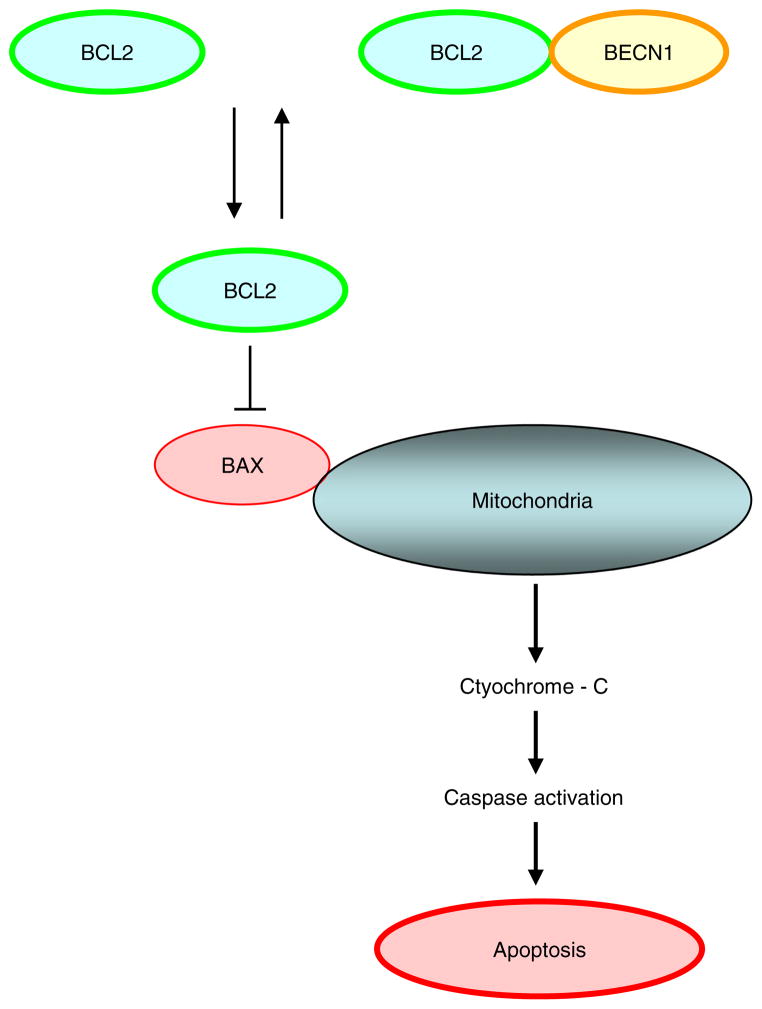
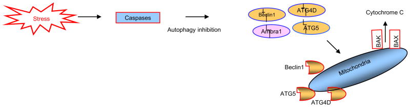
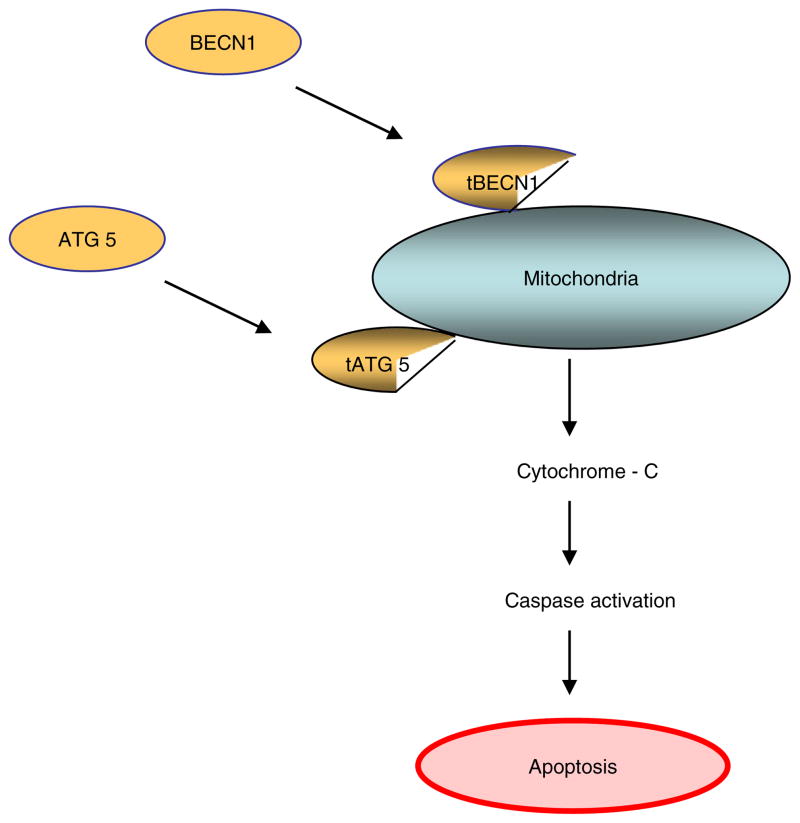
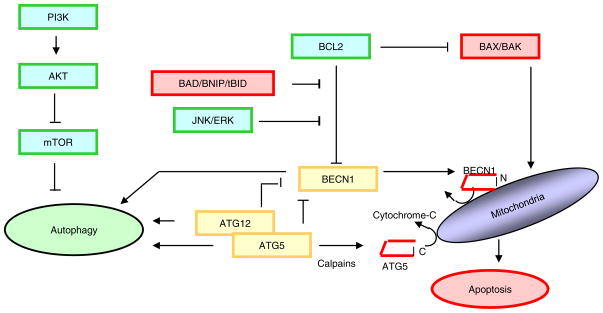
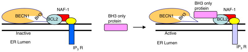
Not surprisingly, the death of a cell is a complex and well controlled process. For several decades, apoptosis, the first genetically programmed death process to be identified has taken centre stage as the principal mechanism of programmed cell death (type I cell death) in mammalian tissues. Apoptosis has been extensively studied and its contribution to the pathogenesis of disease well documented. However, apoptosis does not function alone in determining the fate of a cell. More recently, autophagy, a process in which de novo formed membrane enclosed vesicles engulf and consume cellular components, has been shown to engage in complex interplay with apoptosis. As a result, cell death has been subdivided into the categories apoptosis (Type I), autophagic cell death (Type II), and necrosis (Type III). The boundary between Type I and II cell death is not completely clear and as we will discuss in this review and perhaps a discrete difference does not exist, due to intrinsic factors among different cell types and crosstalk among organelles within each cell type. Apoptosis may begin with autophagy and autophagy can often end with apoptosis, inhibition or a blockade of caspase activity may lead a cell to default into Type II cell death from Type I.
毫不奇怪,细胞的死亡是一个复杂且受到良好控制的过程。几十年来,凋亡作为哺乳动物组织中程序性细胞死亡(I 型细胞死亡)的主要机制,已经成为第一个被确定的具有基因编程的死亡过程的研究焦点。凋亡已经被广泛研究,其对疾病发病机制的贡献也有详细记录。然而,凋亡本身并不能决定细胞的命运。最近,自噬,即新形成的膜封闭小泡吞噬和消耗细胞成分的过程,已被证明与凋亡有复杂的相互作用。因此,细胞死亡已被细分为凋亡(I 型)、自噬性细胞死亡(II 型)和坏死(III 型)。I 型和 II 型细胞死亡之间的界限不是完全清楚的,正如我们将在这篇综述中讨论的,由于不同细胞类型之间的内在因素和每个细胞类型内细胞器之间的串扰,可能不存在明显的区别。凋亡可能始于自噬,自噬通常以凋亡结束,抑制或阻断半胱天冬酶的活性可能导致细胞从 I 型默认进入 II 型细胞死亡。