• 文献检索
  • 文档翻译
  • 深度研究
  • 学术资讯
  • Suppr Zotero 插件Zotero 插件
  • 邀请有礼
  • 套餐&价格
  • 历史记录
应用&插件
Suppr Zotero 插件Zotero 插件浏览器插件Mac 客户端Windows 客户端微信小程序
定价
高级版会员购买积分包购买API积分包
服务
文献检索文档翻译深度研究API 文档MCP 服务
关于我们
关于 Suppr公司介绍联系我们用户协议隐私条款
关注我们

Suppr 超能文献

核心技术专利:CN118964589B侵权必究
粤ICP备2023148730 号-1Suppr @ 2026

文献检索

告别复杂PubMed语法,用中文像聊天一样搜索,搜遍4000万医学文献。AI智能推荐,让科研检索更轻松。

立即免费搜索

文件翻译

保留排版,准确专业,支持PDF/Word/PPT等文件格式,支持 12+语言互译。

免费翻译文档

深度研究

AI帮你快速写综述,25分钟生成高质量综述,智能提取关键信息,辅助科研写作。

立即免费体验

高级弥散成像序列有助于评估局灶性皮质发育不良和癫痫患者。

Advanced diffusion imaging sequences could aid assessing patients with focal cortical dysplasia and epilepsy.

机构信息

Epilepsy Society MRI Unit, Department of Clinical and Experimental Epilepsy, UCL Institute of Neurology, Queen Square, London WC1N 3BG, United Kingdom.

Lysholm Department of Neuroradiology, National Hospital for Neurology and Neurosurgery, University College London Hospitals NHS Foundation Trust, Queen Square, London WC1N 3BG, United Kingdom.

出版信息

Epilepsy Res. 2014 Feb;108(2):336-9. doi: 10.1016/j.eplepsyres.2013.11.004. Epub 2013 Nov 17.

DOI:10.1016/j.eplepsyres.2013.11.004
PMID:24315018
原文链接:https://pmc.ncbi.nlm.nih.gov/articles/PMC3969285/
Abstract

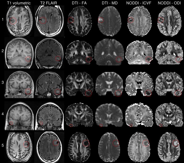
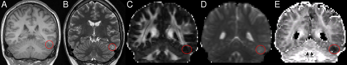
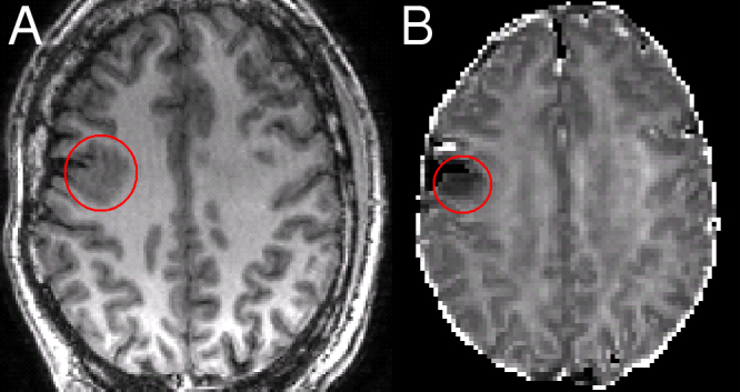
Malformations of cortical development (MCD), particularly focal cortical dysplasia (FCD), are a common cause of refractory epilepsy but are often invisible on structural imaging. NODDI (neurite orientation dispersion and density imaging) is an advanced diffusion imaging technique that provides additional information on tissue microstructure, including intracellular volume fraction (ICVF), a marker of neurite density. We applied this technique in 5 patients with suspected dysplasia to show that the additional parameters are compatible with the underlying disrupted tissue microstructure and could assist in the identification of the affected area. The consistent finding was reduced ICVF in the area of dysplasia. In one patient, an area of reduced ICVF and increased fibre dispersion was identified that was not originally seen on the structural imaging. The focal reduction in ICVF on imaging is compatible with previous iontophoretic data in surgical specimens, was more conspicuous than on other clinical or diffusion images (supported by an increased contrast-to-noise ratio) and more localised than on previous DTI studies. NODDI may therefore assist the clinical identification and localisation of FCD in patients with epilepsy. Future studies will assess this technique in a larger cohort including MRI negative patients.

摘要

皮质发育畸形(MCD),特别是局灶性皮质发育不良(FCD),是难治性癫痫的常见原因,但在结构成像上往往不可见。NODDI(神经丝取向分散和密度成像)是一种先进的扩散成像技术,可提供组织微观结构的额外信息,包括细胞内体积分数(ICVF),这是神经丝密度的标志物。我们将该技术应用于 5 名疑似发育不良的患者,结果表明,额外的参数与潜在的组织微观结构破坏一致,并有助于识别受影响的区域。病变区域的 ICVF 一致减少。在一名患者中,发现了一个原本在结构成像上看不到的区域,其 ICVF 减少,纤维分散增加。成像上的 ICVF 局灶性减少与手术标本中的离子电泳数据一致,比其他临床或扩散图像更明显(支持增加对比度噪声比),比以前的 DTI 研究更局限。因此,NODDI 可能有助于癫痫患者 FCD 的临床识别和定位。未来的研究将在包括 MRI 阴性患者在内的更大队列中评估该技术。

https://cdn.ncbi.nlm.nih.gov/pmc/blobs/a1ef/3969285/3e489681787e/gr2.jpg
https://cdn.ncbi.nlm.nih.gov/pmc/blobs/a1ef/3969285/b173947109d8/mmc1.jpg
https://cdn.ncbi.nlm.nih.gov/pmc/blobs/a1ef/3969285/261c56c651e4/gr1.jpg
https://cdn.ncbi.nlm.nih.gov/pmc/blobs/a1ef/3969285/3e489681787e/gr2.jpg
https://cdn.ncbi.nlm.nih.gov/pmc/blobs/a1ef/3969285/b173947109d8/mmc1.jpg
https://cdn.ncbi.nlm.nih.gov/pmc/blobs/a1ef/3969285/261c56c651e4/gr1.jpg
https://cdn.ncbi.nlm.nih.gov/pmc/blobs/a1ef/3969285/3e489681787e/gr2.jpg

相似文献

1
Advanced diffusion imaging sequences could aid assessing patients with focal cortical dysplasia and epilepsy.高级弥散成像序列有助于评估局灶性皮质发育不良和癫痫患者。
Epilepsy Res. 2014 Feb;108(2):336-9. doi: 10.1016/j.eplepsyres.2013.11.004. Epub 2013 Nov 17.
2
MRI profiling of focal cortical dysplasia using multi-compartment diffusion models.使用多室扩散模型对局灶性皮质发育不良进行 MRI 分析。
Epilepsia. 2020 Mar;61(3):433-444. doi: 10.1111/epi.16451. Epub 2020 Feb 17.
3
Diffusion abnormalities of the corpus callosum in patients with malformations of cortical development and epilepsy.皮质发育畸形和癫痫患者胼胝体的扩散异常
Epilepsy Res. 2014 Nov;108(9):1533-42. doi: 10.1016/j.eplepsyres.2014.08.023. Epub 2014 Sep 16.
4
Application of neurite orientation dispersion and density imaging or diffusion tensor imaging to quantify the severity of cervical spondylotic myelopathy and to assess postoperative neurologic recovery.应用神经丝取向分散和密度成像或弥散张量成像来量化颈椎脊髓病的严重程度,并评估术后神经恢复情况。
Spine J. 2018 Feb;18(2):268-275. doi: 10.1016/j.spinee.2017.07.007. Epub 2017 Jul 12.
5
Neurite orientation dispersion and density imaging parameters may help for the evaluation of epileptogenic tubers in tuberous sclerosis complex patients.神经突方向离散度和密度成像参数可能有助于评估结节性硬化症患者的致痫性结节。
Eur Radiol. 2021 Aug;31(8):5605-5614. doi: 10.1007/s00330-020-07626-7. Epub 2021 Mar 10.
6
Utility and validity of neurite orientation dispersion and density imaging with diffusion tensor imaging to quantify the severity of cervical spondylotic myelopathy and assess postoperative neurological recovery.弥散张量成像的神经丝取向分散和密度成像在定量评估颈椎脊髓病严重程度和评估术后神经恢复中的效用和有效性。
Spine J. 2020 Mar;20(3):417-425. doi: 10.1016/j.spinee.2019.10.019. Epub 2019 Nov 1.
7
The potential role of novel diffusion imaging techniques in the understanding and treatment of epilepsy.新型扩散成像技术在癫痫的理解与治疗中的潜在作用。
Quant Imaging Med Surg. 2015 Apr;5(2):279-87. doi: 10.3978/j.issn.2223-4292.2015.02.03.
8
Neuroimaging in identifying focal cortical dysplasia and prognostic factors in pediatric and adolescent epilepsy surgery.神经影像学在儿科和青少年癫痫手术中识别局灶性皮质发育不良和预后因素。
Epilepsia. 2011 Apr;52(4):722-7. doi: 10.1111/j.1528-1167.2010.02950.x. Epub 2011 Jan 28.
9
Abnormal neurite density and orientation dispersion in unilateral temporal lobe epilepsy detected by advanced diffusion imaging.异常的神经突密度和单侧颞叶癫痫的取向分散通过先进的扩散成像检测到。
Neuroimage Clin. 2018;20:772-782. doi: 10.1016/j.nicl.2018.09.017. Epub 2018 Sep 23.
10
Voxel-based automated detection of focal cortical dysplasia lesions using diffusion tensor imaging and T2-weighted MRI data.使用扩散张量成像和T2加权MRI数据基于体素的局灶性皮质发育不良病变自动检测
Epilepsy Behav. 2018 Jul;84:127-134. doi: 10.1016/j.yebeh.2018.04.005. Epub 2018 May 21.

引用本文的文献

1
Detecting glioblastoma infiltration beyond conventional imaging tumour margins using MTE-NODDI.使用MTE-NODDI检测胶质母细胞瘤超出传统影像肿瘤边界的浸润情况。
Imaging Neurosci (Camb). 2025 Feb 18;3. doi: 10.1162/imag_a_00472. eCollection 2025.
2
Towards precision MRI biomarkers in epilepsy with normative modelling.基于规范建模的癫痫精准MRI生物标志物研究
Brain. 2025 Jul 7;148(7):2247-2261. doi: 10.1093/brain/awaf090.
3
White Matter Integrity Differences in 2-Year-Old Children Treated With ECMO: A Diffusion-Weighted Imaging Study.

本文引用的文献

1
NODDI: practical in vivo neurite orientation dispersion and density imaging of the human brain.NODDI:人脑神经丝取向分散和密度成像的实用体内技术。
Neuroimage. 2012 Jul 16;61(4):1000-16. doi: 10.1016/j.neuroimage.2012.03.072. Epub 2012 Mar 30.
2
Compartment models of the diffusion MR signal in brain white matter: a taxonomy and comparison.脑白质磁共振扩散信号的隔室模型:分类与比较。
Neuroimage. 2012 Feb 1;59(3):2241-54. doi: 10.1016/j.neuroimage.2011.09.081. Epub 2011 Oct 7.
3
The diffusion parameters of the extracellular space are altered in focal cortical dysplasias.
接受体外膜肺氧合治疗的2岁儿童的白质完整性差异:一项扩散加权成像研究。
Eur J Neurosci. 2025 Feb;61(4):e70026. doi: 10.1111/ejn.70026.
4
Gait impairment-related axonal degeneration in Parkinson's disease by neurite orientation dispersion and density imaging.帕金森病中与步态障碍相关的轴突退变:通过神经突方向离散度与密度成像研究
NPJ Parkinsons Dis. 2024 Feb 27;10(1):45. doi: 10.1038/s41531-024-00654-w.
5
Exploring individual fixel-based white matter abnormalities in epilepsy.探索癫痫中基于体素的个体白质异常。
Brain Commun. 2023 Dec 22;6(1):fcad352. doi: 10.1093/braincomms/fcad352. eCollection 2024.
6
C-NODDI: a constrained NODDI model for axonal density and orientation determinations in cerebral white matter.C-NODDI:一种用于确定脑白质中轴突密度和方向的约束性NODDI模型。
Front Neurol. 2023 Aug 3;14:1205426. doi: 10.3389/fneur.2023.1205426. eCollection 2023.
7
Dynamic structural remodeling of the human visual system prompted by bilateral retinal gene therapy.双侧视网膜基因治疗促使人类视觉系统发生动态结构重塑。
Curr Res Neurobiol. 2023 Jun 4;4:100089. doi: 10.1016/j.crneur.2023.100089. eCollection 2023.
8
Multi-tensor diffusion abnormalities of gray matter in an animal model of cortical dysplasia.皮质发育异常动物模型中灰质的多张量扩散异常
Front Neurol. 2023 May 5;14:1124282. doi: 10.3389/fneur.2023.1124282. eCollection 2023.
9
Acquisition and Analysis of Excised Neocortex from Pediatric Patients with Focal Cortical Dysplasia Using Mesoscale Diffusion MRI.使用中尺度扩散磁共振成像对患有局灶性皮质发育异常的儿科患者切除的新皮质进行采集与分析。
Diagnostics (Basel). 2023 Apr 24;13(9):1529. doi: 10.3390/diagnostics13091529.
10
Multimodal combination of neuroimaging methods for localizing the epileptogenic zone in MR-negative epilepsy.多模态神经影像学方法联合用于定位 MRI 阴性癫痫的致痫区。
Sci Rep. 2022 Sep 7;12(1):15158. doi: 10.1038/s41598-022-19121-8.
细胞外间隙的扩散参数在局灶性皮质发育不良中发生改变。
Neurosci Lett. 2011 Jul 15;499(1):19-23. doi: 10.1016/j.neulet.2011.05.023. Epub 2011 May 20.
4
Polymicrogyria includes fusion of the molecular layer and decreased neuronal populations but normal cortical laminar organization.巨脑回畸形包括分子层融合和神经元数量减少,但皮质层结构正常。
J Neuropathol Exp Neurol. 2011 Jun;70(6):438-43. doi: 10.1097/NEN.0b013e31821ccf1c.
5
The clinicopathologic spectrum of focal cortical dysplasias: a consensus classification proposed by an ad hoc Task Force of the ILAE Diagnostic Methods Commission.局灶性皮质发育不良的临床病理谱:国际抗癫痫联盟诊断方法委员会特别工作组提出的共识分类。
Epilepsia. 2011 Jan;52(1):158-74. doi: 10.1111/j.1528-1167.2010.02777.x. Epub 2010 Nov 10.
6
Imaging in the surgical treatment of epilepsy.癫痫的手术治疗中的影像学。
Nat Rev Neurol. 2010 Oct;6(10):537-50. doi: 10.1038/nrneurol.2010.131. Epub 2010 Sep 14.
7
Automated normalized FLAIR imaging in MRI-negative patients with refractory focal epilepsy.MRI阴性的难治性局灶性癫痫患者的自动标准化液体衰减反转恢复序列成像
Epilepsia. 2009 Jun;50(6):1484-90. doi: 10.1111/j.1528-1167.2009.02022.x. Epub 2009 Mar 9.
8
A general framework for experiment design in diffusion MRI and its application in measuring direct tissue-microstructure features.扩散磁共振成像实验设计的通用框架及其在测量直接组织微观结构特征中的应用。
Magn Reson Med. 2008 Aug;60(2):439-48. doi: 10.1002/mrm.21646.
9
Optimal acquisition orders of diffusion-weighted MRI measurements.扩散加权磁共振成像测量的最佳采集顺序
J Magn Reson Imaging. 2007 May;25(5):1051-8. doi: 10.1002/jmri.20905.
10
Seizure outcome after epilepsy surgery in patients with normal preoperative MRI.术前MRI正常的癫痫患者接受癫痫手术后的发作结果。
J Neurol Neurosurg Psychiatry. 2005 May;76(5):710-3. doi: 10.1136/jnnp.2003.026757.