• 文献检索
  • 文档翻译
  • 深度研究
  • 学术资讯
  • Suppr Zotero 插件Zotero 插件
  • 邀请有礼
  • 套餐&价格
  • 历史记录
应用&插件
Suppr Zotero 插件Zotero 插件浏览器插件Mac 客户端Windows 客户端微信小程序
定价
高级版会员购买积分包购买API积分包
服务
文献检索文档翻译深度研究API 文档MCP 服务
关于我们
关于 Suppr公司介绍联系我们用户协议隐私条款
关注我们

Suppr 超能文献

核心技术专利:CN118964589B侵权必究
粤ICP备2023148730 号-1Suppr @ 2026

文献检索

告别复杂PubMed语法,用中文像聊天一样搜索,搜遍4000万医学文献。AI智能推荐,让科研检索更轻松。

立即免费搜索

文件翻译

保留排版,准确专业,支持PDF/Word/PPT等文件格式,支持 12+语言互译。

免费翻译文档

深度研究

AI帮你快速写综述,25分钟生成高质量综述,智能提取关键信息,辅助科研写作。

立即免费体验

胎儿超声心动图技术。

Technique of fetal echocardiography.

作者信息

Lee Mi-Young, Won Hye-Sung

机构信息

Department of Obstetrics and Gynecology, University of Ulsan College of Medicine, Asan Medical Center, Seoul, Korea.

出版信息

Obstet Gynecol Sci. 2013 Jul;56(4):217-26. doi: 10.5468/ogs.2013.56.4.217. Epub 2013 Jul 15.

DOI:10.5468/ogs.2013.56.4.217
PMID:24328006
原文链接:https://pmc.ncbi.nlm.nih.gov/articles/PMC3784141/
Abstract

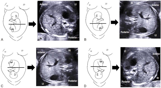
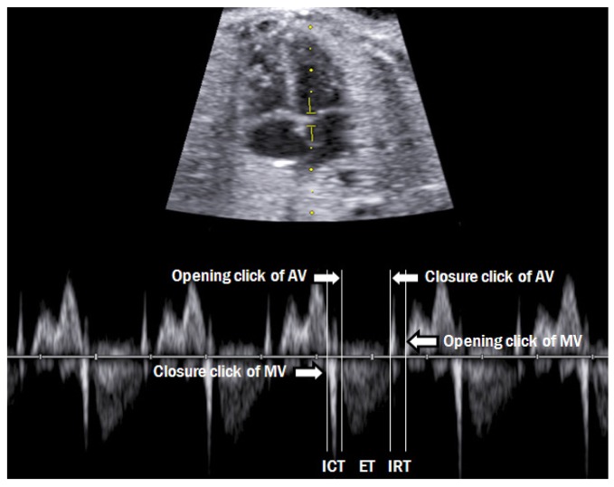
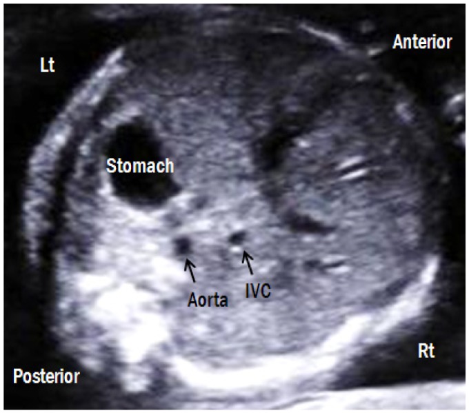
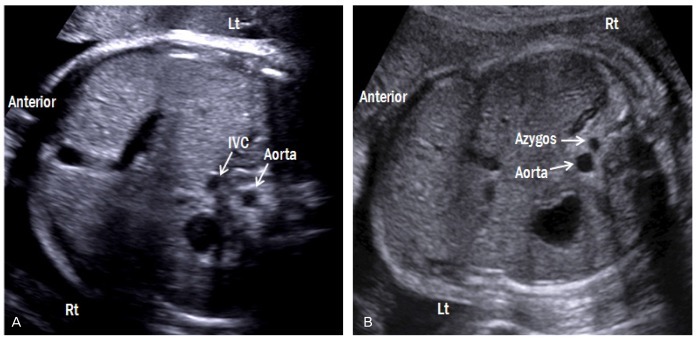
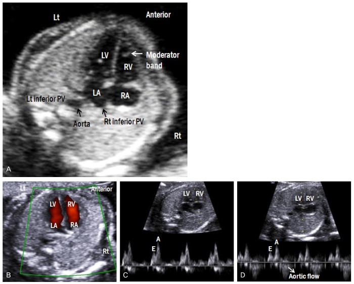
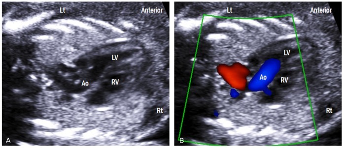
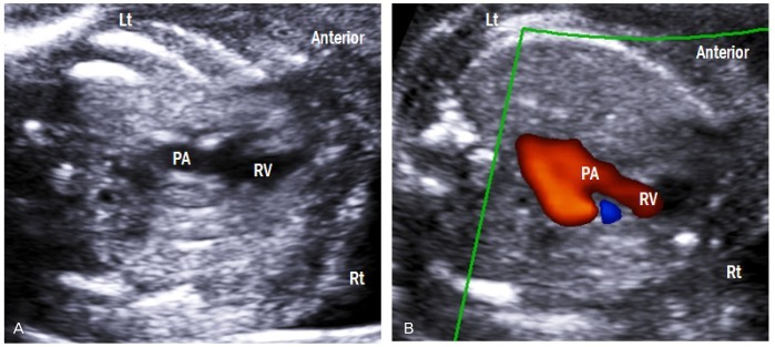
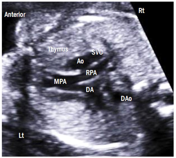
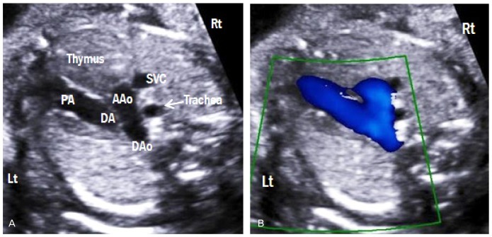
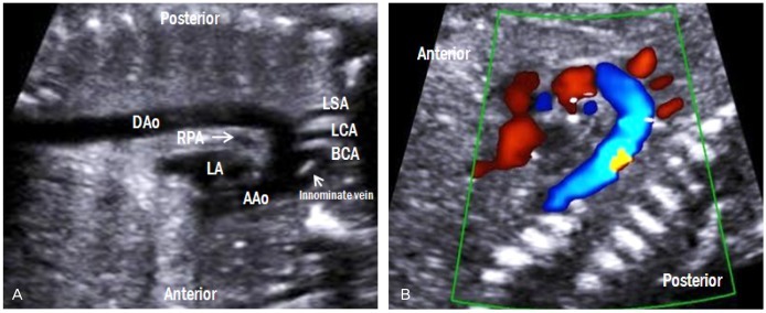
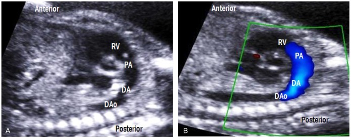
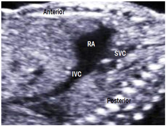
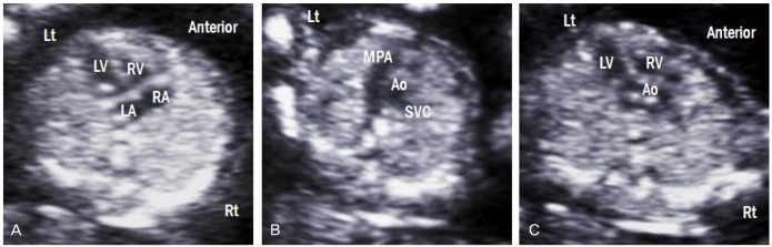
Congenital heart disease is the most common abnormality in the human fetus. Fetal echocardiography has been used to detect the majority of cardiac defects, and it is now part of the routine screening method for fetal evaluation. In this article, we present standard ultrasonographic views of the normal fetal heart obtained during the second trimester, first-trimester fetal echocardiography findings, and a modified myocardial performance index.

摘要

先天性心脏病是人类胎儿中最常见的异常情况。胎儿超声心动图已被用于检测大多数心脏缺陷,并且它现在是胎儿评估常规筛查方法的一部分。在本文中,我们展示了孕中期获得的正常胎儿心脏的标准超声图像、孕早期胎儿超声心动图检查结果以及一种改良的心肌性能指数。

https://cdn.ncbi.nlm.nih.gov/pmc/blobs/7d2a/3784141/100d793ed5ed/ogs-56-217-g014.jpg
https://cdn.ncbi.nlm.nih.gov/pmc/blobs/7d2a/3784141/0af37ed710bc/ogs-56-217-g001.jpg
https://cdn.ncbi.nlm.nih.gov/pmc/blobs/7d2a/3784141/4f2f9b20ebee/ogs-56-217-g002.jpg
https://cdn.ncbi.nlm.nih.gov/pmc/blobs/7d2a/3784141/b1d642a51107/ogs-56-217-g003.jpg
https://cdn.ncbi.nlm.nih.gov/pmc/blobs/7d2a/3784141/49f504194701/ogs-56-217-g004.jpg
https://cdn.ncbi.nlm.nih.gov/pmc/blobs/7d2a/3784141/7baea16d3144/ogs-56-217-g005.jpg
https://cdn.ncbi.nlm.nih.gov/pmc/blobs/7d2a/3784141/79fa2ff55008/ogs-56-217-g006.jpg
https://cdn.ncbi.nlm.nih.gov/pmc/blobs/7d2a/3784141/a1a4102b70de/ogs-56-217-g007.jpg
https://cdn.ncbi.nlm.nih.gov/pmc/blobs/7d2a/3784141/4c7948e75a00/ogs-56-217-g008.jpg
https://cdn.ncbi.nlm.nih.gov/pmc/blobs/7d2a/3784141/f67b313d8bca/ogs-56-217-g009.jpg
https://cdn.ncbi.nlm.nih.gov/pmc/blobs/7d2a/3784141/139b7f44b9cf/ogs-56-217-g010.jpg
https://cdn.ncbi.nlm.nih.gov/pmc/blobs/7d2a/3784141/3d5e00ee2891/ogs-56-217-g011.jpg
https://cdn.ncbi.nlm.nih.gov/pmc/blobs/7d2a/3784141/709be62e70d7/ogs-56-217-g012.jpg
https://cdn.ncbi.nlm.nih.gov/pmc/blobs/7d2a/3784141/276ed885cfea/ogs-56-217-g013.jpg
https://cdn.ncbi.nlm.nih.gov/pmc/blobs/7d2a/3784141/100d793ed5ed/ogs-56-217-g014.jpg

相似文献

1
Technique of fetal echocardiography.胎儿超声心动图技术。
Obstet Gynecol Sci. 2013 Jul;56(4):217-26. doi: 10.5468/ogs.2013.56.4.217. Epub 2013 Jul 15.
2
[Importance of "Guidelines for performing fetal cardiac scan" in prenatal screening for fetal congenital heart disease].《“胎儿心脏扫描操作指南”在胎儿先天性心脏病产前筛查中的重要性》
Zhonghua Fu Chan Ke Za Zhi. 2009 Feb;44(2):103-7.
3
Prenatal diagnosis of congenital heart defects: echocardiography.先天性心脏缺陷的产前诊断:超声心动图检查
Transl Pediatr. 2021 Aug;10(8):2210-2224. doi: 10.21037/tp-20-164.
4
First-trimester transabdominal fetal echocardiography.孕早期经腹胎儿超声心动图检查。
Lancet. 1998 Apr 4;351(9108):1023-7. doi: 10.1016/S0140-6736(97)08406-7.
5
[Accuracy of prenatal diagnosis of congenital heart defects by fetal echocardiography: a 7-year experience in a Chinese tertiary obstetric center].[胎儿超声心动图对先天性心脏病产前诊断的准确性:中国一家三级产科中心的7年经验]
Zhonghua Xin Xue Guan Bing Za Zhi. 2009 Apr;37(4):343-6.
6
Length to width ratio of the ductus venosus in simple screening for fetal congenital heart diseases in the second trimester.孕中期胎儿先天性心脏病简易筛查中静脉导管的长宽比
Medicine (Baltimore). 2016 Sep;95(39):e4928. doi: 10.1097/MD.0000000000004928.
7
Basic heart examination: feasibility study of first-trimester systematic simplified fetal echocardiography.基本心脏检查:孕早期系统性简化胎儿超声心动图的可行性研究
Ultrasound Obstet Gynecol. 2017 Feb;49(2):224-230. doi: 10.1002/uog.15866.
8
Accuracy of fetal echocardiography in the routine detection of congenital heart disease among unselected and low risk populations: a systematic review.胎儿超声心动图在未筛选及低风险人群中常规检测先天性心脏病的准确性:一项系统评价
BJOG. 2005 Jan;112(1):24-30. doi: 10.1111/j.1471-0528.2004.00295.x.
9
Screening for congenital heart disease: a move toward earlier echocardiography.先天性心脏病筛查:向更早进行超声心动图检查迈进。
Am J Perinatol. 2007 Sep;24(8):449-56. doi: 10.1055/s-2007-986681.
10
Early fetal echocardiography.早期胎儿超声心动图。
Birth Defects Res. 2019 May 1;111(8):370-379. doi: 10.1002/bdr2.1414. Epub 2018 Nov 14.

引用本文的文献

1
Longitudinal Behavior of Left-Ventricular Strain in Fetal Growth Restriction.胎儿生长受限中左心室应变的纵向行为
Diagnostics (Basel). 2023 Mar 27;13(7):1252. doi: 10.3390/diagnostics13071252.
2
Application of Artificial Intelligence in Anatomical Structure Recognition of Standard Section of Fetal Heart.人工智能在胎儿心脏标准切面解剖结构识别中的应用。
Comput Math Methods Med. 2023 Jan 24;2023:5650378. doi: 10.1155/2023/5650378. eCollection 2023.
3
Unsupervised Learning-Based Non-Invasive Fetal ECG Muti-Level Signal Quality Assessment.

本文引用的文献

1
Fetal evaluation of the modified-myocardial performance index in pregnancies complicated by diabetes.妊娠期糖尿病胎儿改良心肌做功指数评估。
Prenat Diagn. 2012 Oct;32(10):943-8. doi: 10.1002/pd.3937. Epub 2012 Jul 24.
2
Contribution of the myocardial performance index and aortic isthmus blood flow index to predicting mortality in preterm growth-restricted fetuses.心肌做功指数和主动脉峡部血流指数对预测生长受限早产儿死亡率的作用。
Ultrasound Obstet Gynecol. 2009 Oct;34(4):430-6. doi: 10.1002/uog.7347.
3
Assessment of fetal cardiac function before and after therapy for twin-to-twin transfusion syndrome.
基于无监督学习的非侵入式胎儿心电图多级别信号质量评估
Bioengineering (Basel). 2023 Jan 4;10(1):66. doi: 10.3390/bioengineering10010066.
4
Pregnancy in the time of COVID-19: towards Fetal monitoring 4.0.COVID-19 时期的妊娠:迈向胎儿监测 4.0。
BMC Pregnancy Childbirth. 2023 Jan 16;23(1):33. doi: 10.1186/s12884-023-05349-3.
5
Fetal Cardiac Function: Myocardial Performance Index.胎儿心脏功能:心肌做功指数。
Curr Cardiol Rev. 2022;18(4):e271221199505. doi: 10.2174/1573403X18666211227145856.
6
Variations in ductus arteriosus Doppler parameters in different sonographic views during the second half of gestation.妊娠后半期不同超声检查视图下动脉导管多普勒参数的变化。
Exp Ther Med. 2019 Jan;17(1):502-506. doi: 10.3892/etm.2018.6943. Epub 2018 Nov 8.
7
Clinical characteristics of prenatally diagnosed persistent left superior vena cava in low-risk pregnancies.低风险妊娠中产前诊断的永存左上腔静脉的临床特征
Prenat Diagn. 2016 May;36(5):444-8. doi: 10.1002/pd.4801. Epub 2016 Mar 27.
8
Prenatal diagnosis of atrial isomerism in the Korean population.韩国人群中心房异构的产前诊断。
Obstet Gynecol Sci. 2014 May;57(3):193-200. doi: 10.5468/ogs.2014.57.3.193. Epub 2014 May 15.
Am J Obstet Gynecol. 2009 Apr;200(4):400.e1-7. doi: 10.1016/j.ajog.2009.01.051.
4
Modified myocardial performance index for evaluation of fetal cardiac function in pre-eclampsia.改良心肌性能指数用于评估子痫前期胎儿心功能
Ultrasound Obstet Gynecol. 2009 Jan;33(1):51-7. doi: 10.1002/uog.6272.
5
Clinical implication of isolated right dominant heart in the fetus.胎儿孤立性右优势型心脏的临床意义。
Prenat Diagn. 2007 Aug;27(8):695-8. doi: 10.1002/pd.1756.
6
Z-scores of the fetal aortic isthmus and duct: an aid to assessing arch hypoplasia.胎儿主动脉峡部和动脉导管的Z值:评估主动脉弓发育不全的辅助指标。
Ultrasound Obstet Gynecol. 2007 Jun;29(6):628-33. doi: 10.1002/uog.4021.
7
A modified myocardial performance (Tei) index based on the use of valve clicks improves reproducibility of fetal left cardiac function assessment.基于瓣膜喀喇音使用的改良心肌性能(Tei)指数提高了胎儿左心功能评估的可重复性。
Ultrasound Obstet Gynecol. 2005 Sep;26(3):227-32. doi: 10.1002/uog.1959.
8
Technique of fetal echocardiography.胎儿超声心动图技术
Pediatr Cardiol. 2004 May-Jun;25(3):223-33. doi: 10.1007/s00246-003-0588-y.
9
Clinical impact of first and early second trimester fetal echocardiography on high risk pregnancies.孕早期及孕中期早期胎儿超声心动图对高危妊娠的临床影响。
Heart. 2004 Aug;90(8):921-6. doi: 10.1136/hrt.2003.015065.
10
Early diagnosis of fetal congenital heart disease by transvaginal echocardiography.经阴道超声心动图对胎儿先天性心脏病的早期诊断
Ultrasound Obstet Gynecol. 1993 Sep 1;3(5):310-7. doi: 10.1046/j.1469-0705.1993.03050310.x.